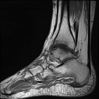
Ankle (Tibiotalar) Osteoarthritis

| |
| Ankle (Tibiotalar) Osteoarthritis | |
|---|---|
| Epidemiology | 3.4% of older adults |
| Causes | Most commonly post-traumatic |
| Risk Factors | Trauma, obesity |
| Clinical Features | Pain and restriction of dorsiflexion and plantarflexion |
| Treatment | weight loss, physical therapy |
There is little research on osteoarthritis of the ankle joint when compared to other sites such as the knee, hip, and hand.
Anatomy
- Main article: Tibiotalar Joint (Talocrural Joint)
The tibiotalar joint, also known as the ankle joint or talocrural joint, is formed by the junction between the distal tibia and fibula and the talus. It has evolved for stability rather than mobility.
Aetiology
Causes are post-traumatic (78%), secondary (13%), and idiopathic (9%)[2]
Post-traumatic cases include patients with a history of ankle trauma or recurrent instability. Of these patients ~50% had a previous malleoli fracture, ~18% a tibial plafond fracture, ~3% a talar fracture, ~6% a tibial shaft fracture, and ~20% ankle ligamentous injuries.
Secondary causes include rheumatoid arthritis, hemochromatosis, haemophilia, clubfoot, osteochondrosis dissecans, and post-infectious arthritis.
Overweight individuals have an increased risk of developing ankle osteoarthritis. Some forms of physical activity may also increase the risk such as ballet dancing.
Epidemiology
It affects approximately 3.4% of older adults in the UK.[3]
Primary osteoarthritis has a mean age of 65.
Post-traumatic osteoarthritis is seen in younger patients with a mean age of 58, but can present in ones early 20s.[2]
Investigations
Plain films: Four weight-bearing standard views:
- Antero-posterior
- Lateral foot
- Mortise view of the ankle
- Specialised hindfoot view (Saltzman view)
There are multiple ankle osteoarthritis classificaiton systems.
MRI: This can help identify early osteochondral lesions.
Clinical Features
Typical symptoms are stiffness, swelling, and aching pain in the ankle joint. The pain is usually activity-related and exacerbated with weightbearing. It is relieved by rest in the early stages. There may be crunching or clicking.
The ankle examination starts with assessing gait. Assess for overall alignment from behind and laterally. The patient is asked to go on tiptoes to view the ankle turning into varus. Assess for swelling surrounding the joint and any skin changes. Palpate the joint lines and surrounding soft tissues. There is usually joint line tenderness. Assess ankle joint motion which is usually limited throughout all planes, but with increased restriction of dorsiflexion and plantarflexion. Restriction of inversion and eversion suggests subtalar joint involvement. Also assess the neurovascular status, and the ipsilateral knee and hip.
Differential Diagnosis
The most important differentials are subtalar joint osteoarthritis, Achilles tendon pathology, and peroneal tendinopathy.
Diffuse
- Ankle (Tibiotalar) Osteoarthritis
- Subtalar Osteoarthritis
- Inflammatory arthropathies - Rheumatoid Arthritis, septic arthritis, osteomyelitis, gout, Osteonecrosis.
- Sarcoid periarthritis
- Benign or malignant neoplasms, myelodysplastic and leukaemic disorders
- Plant-thorn synovitis
- CRPS
Ankle pain persistent following injury
- Fractures - anterior process calcaneus, lateral process talus, posterior process talus (or, rare, os trigonum fracture), osteochondral lesion, tibial plafond chondral lesion, base of fifth metatarsal
- Bony impingements - anterior, posterior, anterolateral
- Atypical sprains - chronic ligamentous instability, medial ligament sprain, syndesmosis sprain (AITFL sprain), subtalar joint sprain
- Tendon injuries - chronic peroneal tendon weakness, peroneal tendon subluxation/rupture, tibialis posterior tendon subluxation/rupture
- Other - inadequate rehabilitation, chronic synovitis, sinus tarsi syndrome
Lateral Ankle Pain
- Peroneal Tendinopathy or recurrent dislocation of peroneal tendons
- Sinus Tarsi Syndrome
- Impingement syndrome (anterolateral, posterior)
- Stress fractures (talus, distal fibula)
- Referred Pain (lumbar, peroneal nerve, superior tibiofibular joint)
- Cuboid Syndrome
- Os Peroneum
- Osteochondritis dissecans (anterior lateral talus)
Medial Ankle Pain
- Tibialis Posterior Tendinopathy
- Flexor Hallucis Longus Tendinopathy
- Medial Calcaneal Nerve Entrapment
- Stress fracture (calcaneal, talar, medial malleolar, navicular)
- Tarsal Tunnel Syndrome
- Posterior Ankle Impingement
- Referred Pain from the lumbar spine
- Complications of acute ankle injuries
- Os tibiale externum (accessory navicular)
- Osteochondritis dissecans (posterior medial talus)
Posterior Ankle Pain
- Midportion Achilles Tendinopathy
- Insertional Achilles Tendinopathy
- Retrocalcaneal bursitis
- Paratenon inflammation
- Posterior impingement syndrome
- Plantaris Tendinopathy
- Sever Disease (Calcaneal Apophysitis)
- Accessory or low soleus muscle
- Referred Pain (neural structures, lumbar spine), and sural neuropathy
- Achilles tendon rupture
- Achilles enthesopathy due to inflammatory arthropathies
- Osteochondritis dissecans (posterior medial talus)
Treatment
Non-operative treatments are physical therapy, weight loss, assistive devices, analgesia, and injections.
With each kilogram of weight loss there is a four-fold reduction in load on the knee joint[4], and it is suspected that the load reduction is even further reduced at the ankle joint.[5]
With using a cane 25% of body weight can be offloaded.
Multiple foot orthoses are available. Rigid hindfoot orthoses provide superior ankle joint motion restriction while still allowing for most forefoot motion.
Corticosteroid injections can provide short term relief.[5]
Hyaluronic acid injections have received a lot of study. There have been four prospective studies, and three RCTs. The prospective studies were promising, but the RCTs were all basically negative.[5]
An RCT of 100 patients with ankle osteoarthritis found that PRP was no better than placebo at 26 weeks for pain and function. This is the first RCT done for the ankle joint.[6]
In those with moderate to severe disease surgery can be considered but surgery. Total ankle arthroplasty is associated with high rates of complications. Ankle arthrodesis severely restricts ankle range of motion.[5]
See Also
References
- ↑ https://radiopaedia.org/articles/osteoarthritis-of-the-ankle
- ↑ 2.0 2.1 Valderrabano V, Horisberger M, Russell I, Dougall H, Hintermann B. Etiology of ankle osteoarthritis. Clin Orthop Relat Res. 2009 Jul;467(7):1800-6. doi: 10.1007/s11999-008-0543-6. Epub 2008 Oct 2. PMID: 18830791; PMCID: PMC2690733.
- ↑ Murray C, Marshall M, Rathod T, Bowen CJ, Menz HB, Roddy E. Population prevalence and distribution of ankle pain and symptomatic radiographic ankle osteoarthritis in community dwelling older adults: A systematic review and cross-sectional study. PLoS One. 2018 Apr 30;13(4):e0193662. doi: 10.1371/journal.pone.0193662. PMID: 29708977; PMCID: PMC5927448.
- ↑ Messier SP, Gutekunst DJ, Davis C, DeVita P. Weight loss reduces knee-joint loads in overweight and obese older adults with knee osteoarthritis. Arthritis Rheum. 2005 Jul;52(7):2026-32. doi: 10.1002/art.21139. PMID: 15986358.
- ↑ 5.0 5.1 5.2 5.3 Khlopas H, Khlopas A, Samuel LT, Ohliger E, Sultan AA, Chughtai M, Mont MA. Current Concepts in Osteoarthritis of the Ankle: Review. Surg Technol Int. 2019 Nov 10;35:280-294. PMID: 31237341.
- ↑ Paget LDA, Reurink G, de Vos RJ, Weir A, Moen MH, Bierma-Zeinstra SMA, Stufkens SAS, Kerkhoffs GMMJ, Tol JL; PRIMA Study Group. Effect of Platelet-Rich Plasma Injections vs Placebo on Ankle Symptoms and Function in Patients With Ankle Osteoarthritis: A Randomized Clinical Trial. JAMA. 2021 Oct 26;326(16):1595-1605. doi: 10.1001/jama.2021.16602. PMID: 34698782; PMCID: PMC8548954.
Literature Review
- Reviews from the last 7 years: review articles, free review articles, systematic reviews, meta-analyses, NCBI Bookshelf
- Articles from all years: PubMed search, Google Scholar search.
- TRIP Database: clinical publications about evidence-based medicine.
- Other Wikis: Radiopaedia, Wikipedia Search, Wikipedia I Feel Lucky, Orthobullets,


