Basic Neurophysiology
Neurons are cells derived from the same embryonic tissue as skin, namely the ectoderm. They are highly differentiated epithelial cells. Their membranes have a unique abilty to transmit electrical signals. The function of the neuron is to provide a rapid transmission of information, which is often over large distances.
Terminology

Nervous System Overview
Basic Nerve Structure

Motor Neuron and CNS Neurones
The nucleus is located in the cell body. Dendrites detect information to deliver to the cell. From the cell body is an extension of the membrane known as the axon that delivers information to a remote site. The axon terminal is called the telodendron. They are typically multipolar neurons.
Pseudounipolar Neurons - Sensory
In the peripheral nervous system, neurons have an analogous structure. In this situation, the cell body is offset from the axon. These cells are designed to reach out into the periphery to receive information. Their distal terminals have a receptor that detect stimuli from peripheral tissues, and is analogous to the dentrite. The axon refers to the entire conducting component of the cell, i.e. everything except the cell body.
Somatic System
The somatic nervous system provides voluntary muscle control and sensory function. Sensory functions are divided into special senses and general senses.
- Special senses: olfaction, vision, hearing, balance, and taste.
- General senses are categorised into:
- Exteroceptors: present in skin and provide nociception (pain, temperature, touch, pressure)
- Interorecptors: present in viscera and provide mechanical and chemical stimuli
- Proprioceptors: present in muscles, joints, and tendons and awareness of posture and movement
The peripheral nervous system is classified into different types of nerve fibres. Different nerve fibres conduct at different velocities depending on their diameter and whether they are myelinated. If a mixed nerve is stimulated mixed activities can be recorded.
There are three peaks of activity, an A wave, a B wave, and a C wave. A waves are further subdivided into α, β, γ, and δ peaks, which are generated by myelinated neurons that are conducting at different velocities. The Aα waves conduct at 120m/s, with progressively slower velocities with the subsequent peaks. The diameter of the A fibres is proportional to their conduction velocity. The C wave is produced by unmyelinated fibres that conduct very slowly. Aα and Aβ are termed large nerve fibres, while Aδ and C fibres are termed small nerve fibres.
- Characteristics of peripheral nerve fibres
| Nerve Fibre | Myelin | Diameter (µm) | Conduction velocity (m/s) | General Function |
|---|---|---|---|---|
| Aα (I) | Yes | 13-20 | 80-120 | Proprioception: muscle spindle primary endings (Ia), golgi tendon organs (Ib), and alpha motor neurons |
| Aβ (II) | Yes | 6-12 | 35-75 | Discriminative sensitivity to mechanical stimuli (touch, vibration), proprioception, pain modulation (block nociceptive information, allodynia in sensitisation) |
| Aγ | Yes | 4-8 | 15-40 | Touch, pressure, and gamma motor neurons. |
| Aδ (III) | Thin | 1-5 | 5-30 | "rapid" pain, crude touch, pressure, temperature. AMH type I for rapid mechanical pain (high heat threshold >53C), AMH type II for rapid heat pain (lower heat threshold 43-47C). |
| B | No | 1-3 | 3-14 | preganglionic autonomic |
| C (IV) | No | 0.2-1.5 | 0.5-2.0 | "second" pain, mechanical, chemical, thermal, pruritis, and postganglionic autonomic. polymodal |
Autonomic Nervous System
The autonomic nervous system differs to the somatic nervous system in that it consists of a two-neuron series, rather than a direct connection. The preganglionic neuron has its cell body in the central nervous system, while the postganglionic neuron has its cell body in the periphery and this innervates the target tissue. The autonomic ganglion is the synaptic connection between the preganglionic and postganglion neurons.
The efferent autonomic system is divided into the sympathetic, parasympathetic, and enteric nervous systems. Preganglionic nerve fibres are myelinated and use acetylcholine as their neurotransmitters. Postganglionic nerve fibres are smaller unmyelinated fibres that use noradrenaline as their neurotransmitters. Sweat glands are an exception and use cholinergic nerves.
Membrane Potential

A nerve cell is able to maintain a membrane potential, abbreviated as Vm. Inside the cell are proteins that have a negative charge, and attract ions such as sodium and potassium. In the cell membrane are sodium and potassium pumps. They expel sodium ions and take up potassium ions. A concentration gradient is formed, with the net effect being that there is a relative positive charge outside the membrane and a negative charge inside the membrane. There is therefore a potential gradient across the membrane, and this measures around -70mV.
There are also channels in the cell membrane that can transmit sodium or potassium. At rest the channels are closed. The gates are able to open allowing sodium to rush into the cell, and potassium to rush outside the cell.
Following the influx of sodium, a generator potential is created. As more sodium moves into the cell, the membrane potential change increases, leading to a greater depolarisation of the membrane. As the amplitude increases, it has a spread of influence along the length of the receptor membrane, this is called electro-tonic spread. The electro-tonic spread eventually reaches the conducting membrane of the neuron.
Channels
In the human body, some ion channels may allow free diffusion, while others are opened by certain events, meaning the channels are gated. So another way that channels can be categorized is on the basis of how they are gated. Although these classes of ion channels are found primarily in the cells of nervous or muscular tissue, they also can be found in the cells of epithelial and connective tissues. The most well studies types of ion channels are the ligand-gated and voltage-gated channels.
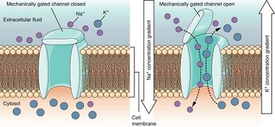
Mechanically Gated Channels When a mechanical change occurs in the surrounding tissue, such as pressure or touch, the channel is physically opened. Thermoreceptors work on a similar principle. When the local tissue temperature changes, the protein reacts by physically opening the channel.
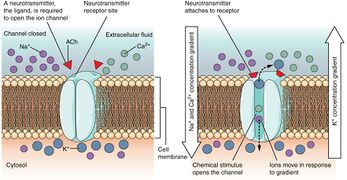
Ligand-Gated Channels When the ligand, in this case the neurotransmitter acetylcholine, binds to a specific location on the extracellular surface of the channel protein, the pore opens to allow select ions through. The ions, in this case, are cations of sodium, calcium, and potassium. Typically found in the post-synaptic membrane.
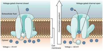
Voltage-Gated Channels Voltage-gated channels open when the transmembrane voltage changes around them. Amino acids in the structure of the protein are sensitive to charge and cause the pore to open to the selected ion (sodium, calcium, etc). Typically found throughout the neuron.
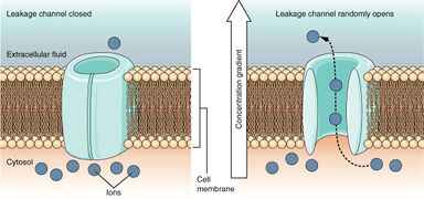
Leakage Channels In certain situations, ions need to move across the membrane randomly. The particular electrical properties of certain cells are modified by the presence of this type of channel.


Nerve Functions
Transduction
This is the ability to detect the stimulus. At the terminal of the nerve fibre is the receptor membrane, which can respond to a variety of stimuli and is able to result in a generator potential through sodium influx. These stimuli can include mechanical, thermal, or chemical influences. Transduction is mediated by specialised receptor ion channels located on the nerve terminal. These receptors determine the threshold and specificity of a sensory neuron's response. Some nociceptors are unimodal which means they are only activated by mechanical, thermal, or chemical stimuli. But most receptors are polymodal, which means they are activated by a variety of stimuli.
Conduction


The resting potential is -70mV and nothing will happen as long as it remains at that level. As the receptor membrane depolarises and the generator potential increases, at a certain point it reaches a threshold value at the conducting membrane. When the threshold value is reached, an action potential is triggered at the conducting membrane.
The action potential is caused by a massive influx of sodium into the cell. This reduces the positivity on the outside and the membrane potential is reduced. Shortly after the potassium conductance is increased, and potassium leaks out of the cell, and this opposes the effect of the sodium conductance, and repolarises the membrane back to normal.
Conduction occurs due to the ability of the nerve membrane to form action potentials. As described, a generator potential is able to initiate an action potential. That action potential itself has a field effect, causing depolarisation of the next section of the membrane through the opening of sodium channels, resulting in a second potential. That second action potential causes a third action potential and so on, and the action potential travels along the axon.
Chemically what is happening is at the site of the action potential, the polarity of the membrane is reversed. Instead of a positive charge outside the membrane and negative charge inside the membrane, there is a negative charge outside and positive charge inside. This charge reversal travel along the membrane.

Certain nerve fibres are myelinated. The myelin sheath is not continuous but rather interrupted at short intervals called the nodes of Ranvier. Myelination allows for saltatory conduction. Instead of action potentials triggering the next most adjacent sodium channels in the membrane, electrical fields stimulate distant sodium channels at the adjacent node of Ranvier. This allows "jumping" of action potentials across the membrane, and this increases the speed of conduction.
Membrane depolarisation is directionally specific under normal circumstances. Once an action potential occurs at a specific site, for a short period that segment of membrane has a refractory period and is unable to respond to electrical information. If the trunk of the nerve is stimulated, action potentials are generated in opposite directions. Antidromic conduction means conduction occurs towards the cell body in the opposite direction of normal, while orthodromic conduction is in the correct direction away from the cell body. And so with trunk stimulation both antidromic and orthodromic conduction occurs.
Transmission

Transmission begins when the action potential reaches the central terminal. There are vesicles in the central end of the axon which contain neurotransmitters. When the action potential arrives at the terminal, the vesicles are pushed into surface of the membrane, and they release the neurotransmitters outside the cell.
| Amines | Amino Acids | Peptides | |
|---|---|---|---|
| Dopamine | GABA | Enkephalins | Substance P |
| Acetylcholine | Glutamate | Cholecystokinin | b-Endorphin |
| Noradrenaline | Glycine | Angiotensin II | CGRP |
| Histamine | Aspartate | Bradykinin | VIP |
| Serotonin | |||
| CGRP: calcitonin gene-related peptide. VIP: vasoactive intestinal peptide | |||
These neurotransmitters may be uniquely released, or there may be several substances released simultaneously. These substances act on the receiving neuron. The dentrites of the receiving neuron contain dentrites that have receptors for these substances. With chemical bonding a miniature potential is generated. The potential is increased with more bonding of neurotransmitters to receptors, eventually causing a depolarisation of the dentrite. This depolarisation spreads across the cell body generating an action potential.
The terminal aspect of the primary neuron is called the pre-synaptic membrane. The receiving membrane is called the post-synaptic membrane. The junction is called the synapse. The communication between these two nerves is chemical not electrical.
Synapses can be chemical or electrical. A chemical synapse provides the ability to control the flow of information. If control isn't required then neither is a synapse. Many nerves can transmit to one receiving nerve. Some of these may be excitatory and other inhibitory. Electrical synapses lack gain, the signal in the postsynaptic neuron is the same or smaller than the presynaptic neuron, but they allow bidirectional flow.
Another important function is the termination of action of transmitters. One method of termination is the natural tendency for neurotransmitters to diffuse away. Another mechanism is the transmitting neuron can re-uptake the neurotransmitters. This allows recycling back into vesicles. A third mechanism is deactivation by enzymes. Enzymes can be found in synapses which changes the nature of the molecule so that it no longer has an effect on the receiving neuron. In the CNS glial cells are able to reuptake neurotransmitters.
The types of neurotransmitters are defined by their substances. Only certain substances can act on certain receptors.
Inhibition of membranes can occur as well as depolarisation through inhibitory neurotransmitters. These inhibit the responsiveness through a process called hyperpolarisation by allowing potassium to leak out of the receiving neuron. The resting potential of the potential, which is normally around -70mV, is increased. This means a stronger stimulus is required to allow depolarisation to the threshold of -55mV.
Resources
See Also
References
- OpenStax textbook

