Baxter's Nerve Entrapment
Baxter's nerve entrapment, also known as Baxter neuropathy, is plantar heel pain arising from compression of the inferior calcaneal nerve (Baxter nerve).
Anatomy

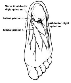
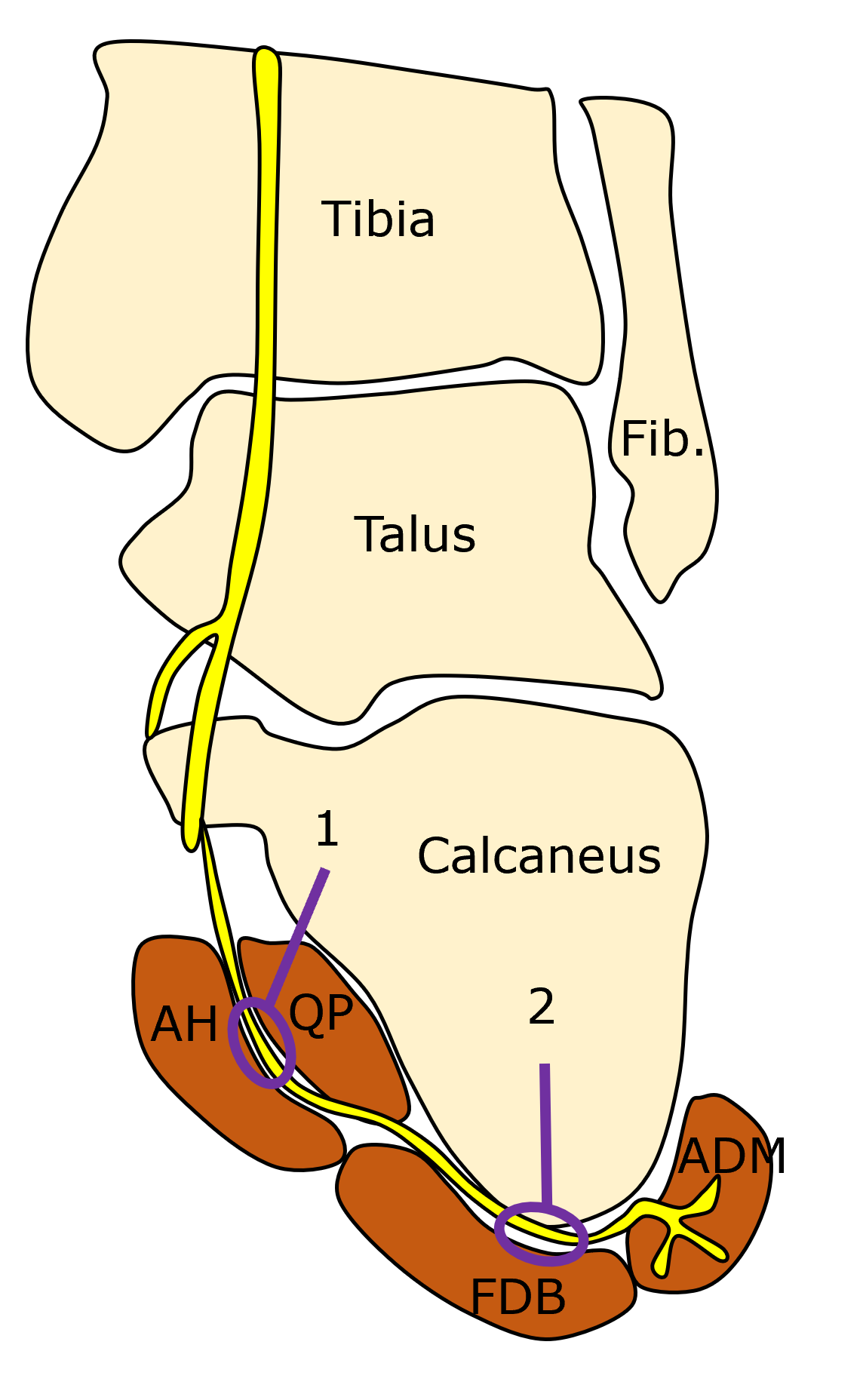
The inferior calcaneal nerve (Baxter nerve) is the first branch of the lateral plantar nerve, which is itself a branch of the tibial nerve. The nerve lies in between the abductor hallucis muscle and quadratus plantae.
The Gray's anatomy image and many other anatomy textbook images are incorrect. The Baxter nerve is quite posterior and lies very close to the calcaneus. It was named after Donald Baxter.[1]
Aetiology
There are three entrapment points:[2]
- Between the deep fascia of the Abductor Hallucis and medial plantar margin of the Quadratus Plantae
- More distally along the anterior aspect of the medial calcaneal tuberosity. A calcaneal plantar enthesophyte and/or soft tissue changes of plantar fasciitis may contribute to entrapment at this second location.
Risk Factors
Obesity, age, diabetes, muscle hypertrophy, hyperpronated foot, pes planus, plantar calcaneal spur, plantar fasciitis, spondyloarthritis.
Clinical Features
The clinical features may be similar to or coexist with plantar fasciitis. However there may be less morning pain, occasional altered sensation, and intrinsic muscle atrophy.
Differential Diagnosis
- Plantar Fasciitis
- Fat Pad Contusion
- Calcaneal fractures (traumatic and stress)
- Inferior Calcaneal (Baxter) Nerve Entrapment
- Medial Calcaneal Nerve Entrapment
- Lateral Plantar Nerve Entrapment
- Medial Plantar Nerve Entrapment
- Tarsal Tunnel Syndrome
- Lumbar Radicular Pain
- Talar stress fracture
- Retrocalcaneal bursitis
- Spondyloarthritis
- Osteoid osteoma
- CRPS
Investigations

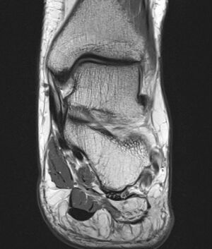
On MRI in the acute phase there is decreased T1 signal intensity and increased T2 signal intensity with fat saturation in the muscles innervated by the Baxter nerve. In the chronic phase there is fatty change of the abductor digiti minimi muscle, and occasionally the Flexor Digitorum Brevis and Quadratus Plantae.
Treatment
The nerve can be injected with corticosteroid and/or hydrodissected under ultrasound guidance.
External Links
Baxter's Nerve (First Branch of the Lateral Plantar Nerve) Impingement - Radsource
References
- ↑ 1.0 1.1 Baxter, D. E.; Thigpen, C. M. (1984-07). "Heel pain--operative results". Foot & Ankle. 5 (1): 16–25. doi:10.1177/107110078400500103. ISSN 0198-0211. PMID 6479759. Check date values in:
|date=(help) - ↑ Moroni, Simone; Zwierzina, Marit; Starke, Vasco; Moriggl, Bernhard; Montesi, Ferruccio; Konschake, Marko (2019-01). "Clinical-anatomic mapping of the tarsal tunnel with regard to Baxter's neuropathy in recalcitrant heel pain syndrome: part I". Surgical and radiologic anatomy: SRA. 41 (1): 29–41. doi:10.1007/s00276-018-2124-z. ISSN 1279-8517. PMC 6514163. PMID 30368565. no-break space character in
|title=at position 123 (help); Check date values in:|date=(help) - ↑ Bauones, S., Feger, J. Baxter neuropathy. Reference article, Radiopaedia.org. (accessed on 15 Apr 2022) https://doi.org/10.53347/rID-25994
- ↑ Baxter's nerve Sd. Procedure: corticosteroid injection & orthotics. Good result. @Dr_Ramon_Balius #MSKUltrasound pic.twitter.com/ED9RcmqJnz
Literature Review
- Reviews from the last 7 years: review articles, free review articles, systematic reviews, meta-analyses, NCBI Bookshelf
- Articles from all years: PubMed search, Google Scholar search.
- TRIP Database: clinical publications about evidence-based medicine.
- Other Wikis: Radiopaedia, Wikipedia Search, Wikipedia I Feel Lucky, Orthobullets,


