Carpal Instability
Anatomy
The carpal bones give stability to the wrist in various positions. The lunate plays a key role in carpal stability. The lunate acts as the main both from which the other bones move around.
Ligaments
Intrinsic Ligaments
The interosseous ligaments connect the carpal bones.
- ScaphoLunate ligament (SLL). Three portions, dorsal is the strongest, the proximal/membranous is associated with the capsule, and the palmar.
- LunoTriquetral ligament (LTL)
- ScaphoTrapezioTrapezoid ligament (STT)
Extrinsic Ligaments
The extrinsic ligaments connect the radius to the carpus.
- Volar RadioScaphoCapitate ligament
- Volar RadioLunoTriquetral ligament
- Volar UlnoLunate
- Volar UlnoTriquetral
- Volar RadioUlnar
- Dorsal RadioTriquetral
- Dorsal RadioUlnar
TFCC
Thumb Ligaments
- Superficial Anterior Oblique Ligament
- Deep Anterior Oblique Ligament
- Dorsal Radial Ligament
Kinematics
Rows and Columns
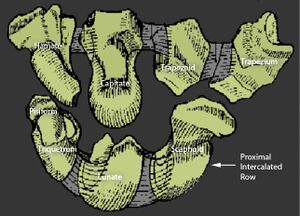
There are two rows and three columns
The two rows are termed proximal and distal. The scaphoid acts as a bridging bone and translates some of the movements from the proximal to distal row in giving more stability to radial and ulnar deviations.. There is motion within and between rows.
There are three columns. On the radial side is the lateral column and this contains the scaphoid, trapezoid, and trapezium; it provides longitudinal stability. On the ulnar side is the medial column and this contains the triquetrum and provides rotational stability. The lateral and medial columns move to accommodate ulnar and radial deviation. The central column contains the lunate, capitate, and hamate; it doesn't deviate a lot, rather there is movement around it.
The centre of rotation happens through the lunocapitate joint. The radial column takes about 75% of load, while the ulnar column takes about 25% of load.
Radial and Ulnar Deviation
Radial deviation: the scaphoid flexes forcing the lunate into palmar flexion, the hamate rotates into "high" position.
Ulnar deviation: scaphoid extends (it fills the anatomical snuff box) pulling the lunate into dorsiflexion hamate rotates into "low" position.
Aetiology
- Trauma
- Sequelae of trauma
- Degenerative
- Secondary for example osteonecrosis
Pathophysiology
The lunate is in a state of dynamic balance. As long as the scapholunate and lunotriquetral ligaments are intact, and there are no other problems in the distal radius, the lunate sits in the middle and doesn't move in a dorsal or volar direction. All the forces are balanced.
When the scaphoid flexes it tries to pull the lunate towards the volar side. When this happens the lunotriquetral joint on the other side stops it from going down. The triquetrum tries to extend to keep the lunate in a balanced position.
If the balance is interrupted, the lunate follows the unbalanced force. See video.
Carpal instability can be defined as the inability of the wrist to maintain a normal balance between the articulating surfaces under physiologic loads (dyskinetics) and/or movements (dyskinematics)
Classification
Carpal instability is called dissociative if it is happening in between the carpal bones in the same row. It is called non-dissociative when it is happening between the two rows. If it is affecting both rows and the columns it is called complex. If it is happening due to non-carpal problems it is called adaptive. CI refers to carpal instability.
- CID (dissociative): DISI and VISI
- CIND (non-dissociative): radiocarpal, midcarpal, ulnar translocation
- CIC (complex): perilunate dislocation
- CIA (adaptive): instability not in carpal, malunited wrist fracture
The deformity can be dynamic or static. This is why it is important to take loading radiograph views (clenched fist, radial and ulnar deviation). A static MRI can incorrectly grade an injury without these dynamic x-ray views.
CID predominantly happens in the proximal row. It can happen in the distal row but it is very rare. DISI is more common than VISI. DISI stands for dorsal intercalated segment instability, is due to scapholunate ligament instability, and the scapholunate angle is increased.
Clinical Features
The features to look for are swelling, reduced range of motion, localised tenderness, neuropathies, and deformities.
The astute examiner with a good knowledge of surface landmarks may find localised tenderness over an area of relevant ligamentous disruption. For scapholunate tenderness, first find the Listers tubercle dorsally and go exactly distal to it. For lunotriquetral tenderness, first find the radioulnar joint, and go just distal to it.
Look for signs of gross deformities, and evidence of previous operations. Previous operative history is important, for example a scapholunate disruption could have been missed at the time of a distal radius fracture repair, or there may have been a previous non-union.
The range of motion is usually reduced.
Common Pitfalls
One pitfall is failure to accurately interpret the history from the patient. The patient may say that they feel like a bone has moved out of their wrist . The examiner then sees a normal x-ray and assumes everything is fine and incorrectly diagnose wrist sprain. The x-ray is normal in 25% of cases of lunate dislocation, and often the x-ray finding are quite subtle.
Imaging
Radiographs
- AP, lateral, oblique
- Clenched fist
- Radial and ulnar deviation - useful for detecting intrinsic joint issues
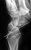
The angles are the ScaphoLunate angle, RadioLunate angle, RadioScaphoid angle, and CapitoLunate angle.
The most important angle is the ScaphoLunate angle.
To measure the SL angle, draw a line between the inferior margin of the scaphoid tuberosity and body of the scaphoid, draw another line perpendicular to the lunate. The angle between these is the SL angle. The normal range is 30-60 degrees. Anything less than 30 is called VISI, anything more than 60 is called DISI.
DISI x-ray findings
- AP View
- Terry Thomas Sign: Scapholunate gap >3mm with clenched fist view on the AP view.
- Cortical ring sign: caused by scaphoid malalignment.
- Humpack deformity: with DISI associated with an unstable scaphoid fracture
- Scaphoid shortening
- Lateral view
- SL angle more than 70 degrees
Other
Arthrogram
MRI and MRI arthrogram
Arthroscopy is the gold standard.
Treatment
Nonoperative treatment is a controversial area.
If there is a fracture it should be fixed. For malunion, address the cause if no ligaments are affected. Percutaneous K wires can be used. Scapholunate repair is the preferred treatment for acute, subacute or controversially for chronic reducible. Scapholunate reconstruction is for chronic or poor ligament quality. Fusion for chronic and degenerative joint disease.


