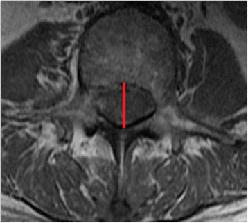
Congenital Lumbar Spinal Stenosis

| |
| Congenital Lumbar Spinal Stenosis | |
|---|---|
| Synonym | Developmental Lumbar Spinal Stenosis, Short Pedicle Syndrome. |
| Definition | No uniform definition |
| Pathophysiology | Abnormal development of the dorsal spinal elements |
| Clinical Features | Similar to other types of spinal stenosis with neurogenic claudication |
| Tests | MRI showing reduced mid-sagittal spinal canal diameter (definitions vary) |
| DDX | Degenerative lumbar spinal stenosis |
| Treatment | Conservative, multi-level spinal instrumentation. |
The incidence of CLSS is unknown. Cheung et al did MRI imaging on 66 patients awaiting surgery for spinal stenosis, and 82 asymptomatic subjects. All were of Chinese ethnicity. All 66 subjects in the patient group had DSS while none of the 82 asymptomatic subjects had developmental canal narrowing.[1] Affected individuals tend to get symptoms in the fourth and fifth decades of life. This is contrasted to degenerative lumbar spinal stenosis where symptoms usually develop after the sixth decade of life.[2]
Aetiopathogenesis
CLSS is thought to be due to abnormal development of the dorsal spinal elements that occurs after birth, with the canal narrowing being primarily a result of the decreased pedicle length.[3] The aetiology is unknown, except for individuals with achondroplasia. CLSS is different to degenerative lumbar spinal stenosis, as the stenosis is not limited to only one or two segments, but rather uniformly affects the entire lumbar spine. Individuals with CLSS are susceptible to even minimal degenerative changes (such as a bulging disc, or apophyseal ring separation) that can compromise the already narrowed spinal canal and cause clinical symptoms.[2] It is possible that patients with degenerative lumbar spinal stenosis presenting later in age have a pre-existing developmentally narrrow canal.[1] [4]
Development of the spinal canal begins early in life. Up to 14 weeks of gestation, the area of the spinal canal is similar throughout all levels from the lumbar and sacral spine. After 30 weeks, the upper lumbar segments grow faster than the lower lumbar spine. At birth, L5 is only 50% mature, with the remaining growth occurring over the first five years of life. By one year of age, the curved surface area (CSA) of the canal at L3 and L4 are adult sized. L1–L4 are 70% of adult size at birth, with the remaining 30% growth being achieved during the first postpartum year. Considering the intrauterine development, patients with predominant L1-L4 involvement may have had an intrauterine insult or growth impairment. While L5 has more potential to catch up in growth. Growth impairment during early infancy does not tend to affect the upper lumbar region as it has achieved most of its growth in utero. L5-S1 involvement could indicate growth impairment over the first 5 years of life.[4]
Definition
There is no uniform definition of CLSS in the literature. Many studies suggest mid-sagittal spinal canal diameter values between 10 and 17mm and don't clarify how many levels it needs to be documented on. Other authors use the cross-sectional area, but this is not practical for everyday use. Soldatos et al used 14mm on at least one level as a cutoff, which they thought "summated" previous reports.[2] Cheung et al used the midline AP bony canal diameter, and had different values for each segment. (L1 <20 mm, L2 <19 mm, L3 <19 mm, L4 <17 mm, L5 <16 mm, S1 <16 mm).[1]
Some but not all authors differentiate developmental from congenital lumbar spinal stenosis. Kitab states that congenital anatomic changes or malformations (eg, an excessive scoliotic or lordotic curve) invariably characterises the congenital type.[4]
Clinical Features
Patients may have a similar clinical presentation to other types of stenosis. The clinical hallmark finding of lumbar stenosis is neurogenic claudication (affecting 91%). This presents as intermittent pain or paraesthesias in the legs that comes on with walking and standing, and is classically relieved with spinal flexion. Back pain is very common affecting 95% of individuals. Weakness (33%), and voiding disturbances (12%) may also occur.[3]
Imaging
On MRI, there is a reduced mid-sagittal spinal canal diameter (definitions vary). There is an increased incidence of circumferential and shallow annular bulges, foraminal and anterior disc herniations, annular tears, and spondylolisthesis. They found that the AP bony canal diameter in CLSS gradually decreased from cranial to caudal, where in control subjects the diameter was uniform.[1]
Kitab et al found that global pathology and multilevel involvement with L3, L4, and L5 segments are involved more commonly and severely, whereas severe stenosis, at L1, L2, and S1 occurs infrequently. There are three spinal canal morphologies in CLSS group:[4]
- “Flattened” canal with predominantly reduced spinal canal AP diameter and flattened spinal canal contents
- Predominantly reduced interlaminar angle and consequent prominence of the anteromedial border of facets
- Combined type, with global reduction of all canal parameters, including the AP and transverse diameters, and the interlaminar angle.
The authors also compared CLSS patients to controls who had an MRI for an acute back pain episode, and found that there was no significant difference in the incidence of degenerative disc[4]
Treatment
Due to CLSS generally involving the entire lumbar spine, surgical treatment commonly required multi-level intervention. Compare this to treatment for degenerative lumbar spinal stenosis which usually only needs focal intervention.[2]
References
- ↑ 1.0 1.1 1.2 1.3 Cheung et al.. Radiographic indices for lumbar developmental spinal stenosis. Scoliosis and spinal disorders 2017. 12:3. PMID: 28239663. DOI. Full Text.
- ↑ 2.0 2.1 2.2 2.3 Soldatos et al.. Spectrum of magnetic resonance imaging findings in congenital lumbar spinal stenosis. World journal of clinical cases 2014. 2:883-7. PMID: 25516864. DOI. Full Text.
- ↑ 3.0 3.1 Singh et al.. Congenital lumbar spinal stenosis: a prospective, control-matched, cohort radiographic analysis. The spine journal : official journal of the North American Spine Society 2005. 5:615-22. PMID: 16291100. DOI.
- ↑ 4.0 4.1 4.2 4.3 4.4 Kitab et al.. Anatomic radiological variations in developmental lumbar spinal stenosis: a prospective, control-matched comparative analysis. The spine journal : official journal of the North American Spine Society 2014. 14:808-15. PMID: 24314904. DOI.
Literature Review
- Reviews from the last 7 years: review articles, free review articles, systematic reviews, meta-analyses, NCBI Bookshelf
- Articles from all years: PubMed search, Google Scholar search.
- TRIP Database: clinical publications about evidence-based medicine.
- Other Wikis: Radiopaedia, Wikipedia Search, Wikipedia I Feel Lucky, Orthobullets,


