Diffuse Idiopathic Skeletal Hyperostosis

| |
| Diffuse Idiopathic Skeletal Hyperostosis | |
|---|---|
| Synonym | DISH, ankylosing hyperostosis, Forestier disease, Forestier-Rotes-Querol disease |
| Epidemiology | Varies across populations. |
| Causes | Unknown |
| Classification | Niwayama and Utsinger, and Resnick for research purposes |
| Risk Factors | Insulin resistance, demographic variations, age |
| History | May or may not have pain |
| Examination | Restricted spinal range of motion particularly lateral flexion |
| Diagnosis | Radiographic, calcification of soft tissues mainly ligaments and entheses, with spinal particularly thoracic predilection |
| Tests | Radiograph, CT |
| DDX | Degenerative spine disease, ankylosing spondylitis, ossification of the posterior longitudinal ligament. |
| Treatment | Supportive, surgical management in certain cases of cervical spine involvement |
| Prognosis | Generally good, may cause reduced spinal mobility. Rare potentially catastrophic sequelae from cervical spine disease. |
Diffuse idiopathic skeletal hyperostosis (DISH) is a progressive noninflammatory condition causing calcification and ossification of ligaments and entheses with a spinal predominance. The condition may be asymptomatic, or may be associated with spinal pain and restricted movement.[1]
Aetiology
The cause of DISH is unknown, however it has been thought that certain factors may be important in its development. These include mechanical factors, diet, drugs, environmental exposures, and metabolic conditions. It is thought that there is abnormal osteoblastic differentiation and activity located at the entheses, as some patients may be deficient in certain inhibitors of bone formation such as Dickkopf-1 and matrix Gla protein.[1]
- Mechanical factors[1]
A right-sided prominence of bony bridging is seen at the thoracic spine. There may be mechanical factors associated with the location of the aorta on the left hand side, perhaps protecting that side. Interestingly, in those with dextrocardia or situs inversus, the bony bridging is more prominent on the left.
Mechanical forces may affect bone formation. In those with ossification of the posterior longitudinal ligament (may occur with DISH or independently), ligamentous stretching can increase prostaglandin I2 synthase which results in stimulation of osteogenic differentation. Dickkopf-1, an inhibitor of osteoblastogenesis, may be reduced in those with DISH thereby promoting hyperostosis.
There does not appear to be a difference in the amount of heavy work done in those with DISH compared to controls.
- Environmental factors and diet[1]
Fluoride has been investigated but found unlikely to be a cause. There are mixed reports of the association between higher serum retinol (signifying excessive vitamin A exposure) levels and DISH.
- Medications[1]
Isotretinoin, which is a derivative of vitamin A, and other retinoids are associated with hyperostosis, but more-so in extraspinal areas.
- Metabolic conditions[1]
It is thought that metabolic factors many contribute to increased osteoblastic activity in DISH. Some studies report increased levels of insulin, insulin-like growth factor 1, and growth hormone. Insulin-like growth factor 1 stimulates osteoblasts, and growth hormone can increase the production of this factor.
There are some intriguing demographic studies looking at the association between DISH and metabolic conditions. The Pima Native American people have both a high incidence of DISH and metabolic conditions such as obesity, hypertension, diabetes mellitus, and hyperinsuliaemia. DISH appears to be more common in obese Caucasian people, and possibly also in those with diabetes mellitus. DISH is also seen in up to 20 percent in acromegalic patients.
Also seen in the condition is increased vascularity of the ossified ligaments. It is not known whether this is a cause or a downstream effect of DISH.
Other factors have been studied in their role in DISH. Platelet-derived growth factor-BB and transforming growth factor-1-beta in the cells of ligaments can increase levels of nuclear factor kappa B. Nuclear factor kappa B can promote osteoblastogenesis. Curiously, there are elevated levels of matrix Gla in DISH, but this protein is thought to inhibit bone formation.
Epidemiology
DISH is in general more common in men than woman, and it is rare under the age of 40. There are some demographic variations. Pacific islanders have very high rates of DISH, one study in South Auckland found a rate of 44% on a retrospective review of chest xrays in those over 50 as assessed by Resnik criteria which is criteria for end stage disease, with negligible sex differences. Maori were at 38%, and European at 22%.[2]. American blacks have lower levels of DISH than whites. The Pima Native American people can develop DISH young, with rates rising to 48% in men and 12% in women in those over 55 years old. In Finland the prevalence is 3.8% in men and 2.6% in women in those over 40. In Hungary the prevalence is 4.9% in men and 1.4% in women over age 50. In Japan, ossification of the posterior longitudinal ligament, mainly in the cervical spine, is 80 time more common than in Europe at 4%. It is important to note that studies done in hospitals find a higher prevalence of DISH than the general population. [1]
Clinical Features

Spinal involvement DISH has traditionally been thought of as a pure radiographic condition. This is often true but DISH can cause marked reductions in spinal mobility, and some patients have spinal pain. [3] Postural abnormalities may resemble that of advanced ankylosing spondylitis.[4] Patients with DISH may have pain in the neck, thoracic spine, low back, and/or the extremities. Thoracic spine involvement is present universally in advanced cases, and typically involves T7-T11. There may be reduced spinal mobility. Myelopathy has been reported to occur in some individuals. Dysphagia and airway obstruction can also occur. It is unclear whether it is associated with other musculoskeletal or systemic diseases. Most studies on clinical features are done in patients seen in clinics and the hospital, and so are more likely to have symptoms.[1]
- Musculoskeletal manifestations[1]
Morning spinal stiffness is present in around 80% of individuals, which is a symptoms normally associated with inflammatory conditions. Higher pain levels are seen in those with DISH compared to lumbar spondylosis and healthy controls. Disability scores are also higher in DISH compared to healthy controls, and patients may reports difficulty bending and having weak grip strength. Men are less likely to complete five chair stands without using their arms.
Radicular pain and lumbar spinal stenosis are not a typical features of DISH, and if present it may be unrelated.
Pain in the thoracic spine is the most common symptoms and occurs in 40 to 80% of subjects. Pain in the extremities and dysphagia are less common, reported in 15 to 25%. Stridor is a rare symptom resulting from large C2 to C3 anterior osteophytes. This may cause odynophagia and otalgia due to hypopharyngeal ulceration from the pressure. There is an increased risk of dysphagia, due to compression of the oesophagus from anterior cervical osteophytes. Other manifestations of cervical disease are hoarseness, aspiration pneumonia, sleep apnoea, atlantoaxial subluxation or pseudoarthrosis, and thoracic outlet syndrome.
There are mixed reports on the association between radiologic findings and symptoms at that site. There appears to be a significant association between shoulder hyperostosis and shoulder symptoms, as well as a non-significant association between elbow hyperostosis and elbow symptoms. Meanwhile there is no apparent link between spinal hyperostosis and spinal pain. In those with DISH and chronic low back pain, there is also a non-significant increase in media epicondylitis, knee enthesitis, plantar fasciitis, and dysphagia, compared to patients with "lumbar spondylosis."
Chalk stick fractures, also known as carrot stick fractures (due to the similar appearance to broken chalk sticks and carrots), can occur which are fractures of the fused spine, and are four times more common in the ankylosed spine. This classically occurs with ankylosing spondylitis but can occur in DISH and OPLL, too. The fracture normally occurs at the disco-vertebral junction in the upper thoracic or lower cervical spine.
When several contiguous segments of the spine are fused, the fused column acts as a lever arm. This places greater than normal stresses on the spine. These fractures often occur following minimal trauma due to the altered biomechanics of the spine.
- Neurological manifestations[1]
Ossification of the posterior longitudinal ligament (OPLL) can cause spinal cord compression and myelopathic symptoms. This appears to be more common in Asian people. Some patients have developed tetraparesis and tetraplegia. Symptoms of cervical myelopathy development that should alert the clinican are a sharp, shoorting neck pain, sudden loss of neck range of motion, unsteady gait and hyperreflexia, and new extremity sensory symptoms.
Other rare findings are horner's syndrome, recurrent laryngeal nerve palsy, and vertebral artery insufficiency.
Examination
Physical examination may reveal decreased spinal range of motion, especially in the thoracic spine. Typically there is a loss of thoracic lateral flexion. There may be nodules palpable at the enthesis. The most common sites of these nodules are the Achilles, knee, and elbow. There appears to be an increased rate of Bouchard and Heberden nodes in those with DISH.[1]
Radiological findings
Radiological manifestations are the hallmark of DISH, characterised by ossification of the paravertebral ligaments and peripheral entheses. CT is generally more sensitive than radiographs.[1]
Spinal Inolvement
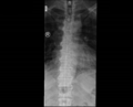
Thoracic DISH: Confluent ossification of multiple contiguous vertebral bodies
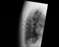
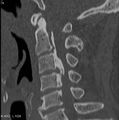
Thoracic DISH: "Melted candle wax" appearance of calcification and ossification, note right side predominance.
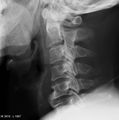
C3-C5 osteophytes causing dysphagia.
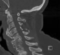
OPLL: case 1 on plain film
OPLL: case 1 on CT


See articles on radiopaedia.org for copious examples of DISH and OPLL
The thoracic spine is the most commonly affected, but there may also be abnormalities in the cervical spine, lumbosacral spine, and extraspinal locations. Full-flowing osteophytosis appears to take 10 years to form.[5] There is characteristic "flowing mantles" of ossification at the anterior longitudinal ligament, and to a lesser degree at the paravertebral connective tissue and the peripheral annulus fibrosis. Hyperostosis may be present in one or more sections of the spine. The anterior longitudinal ligament covers the anterior and anterolateral part of the spine and so AP radiographs can be misleading by only seeing lateral ossifications. [3]
In addition to anterior anterior longitudinal ligament, there may be syndesmosis ossification of the ligamenta flava, superaspinous, and interspinous ligaments. Enthesopathies may also occur and together with syndesmosis ossification can cause spinal canal stenosis. In the cervical spine ossification of the posterior longitudinal ligament (OPLL) can also cause stenosis.[1]
- Thoracic spine
There is flowing linear calcification and ossification along the anterior and right anterolateral vertebral bodies, continuing along the disc space, involving at least 4 contiguous vertebrae. There is a lucent area between the anterior longitudinal ligament and the mid-portion of the vertebral body. Right sided involvement is preponderant.[1]
- Cervical spine
Hyperostosis is first seen along the anterior vertebral body, especially the inferior lip of the vertebra. A downward-pointing spur may be seen. The lower cervical spine is the most commonly affected. The hyperostosis is often symmetrical, and may cause displacement of the trachea and oesophagus. Ossifications are nonflowing on lateral imaging unlike the thoracic spine. Disc narrowing may be present. OPLL is less common than ossification of the anterior longitudinal ligament.[1]
- Lumbar spine
Radiographic findings are similar to the cervical spine. However the spurs tend to point upwards. Flowing ossification is less common than the thoracic spine. There is symmetrical involvement. Complete bridging is uncommon. Disc narrowing may be present. OPLL is less common than in the cervical spine. [1]
- Sacroiliac joints
There may be changes that can be confused with ankylosing spondylitis. The upper portion of the sacroiliac joint is the ligamentous area and may show vacuum phenomenon, narrowing, sclerosis, or ankylosis. The lower two thirds of the joint is the synovial section and does not show changes. Joint capsule ossification can occur of the anterior joint which can resemble changes seen in postinflammatory AS falsely looking like obliteration of the sacroiliac joints. CT can be used to differentiate sacroiliac joint changes.[3]
- Facet joints
There may be narrowing and hypertrophic changes with capsular ossification. There is no ankylosis of facet joints in DISH. Similar changes can be seen in the costovertebral and costotransverse joints manifesting as reduced chest expansion in advanced DISH.[3]
- Ossification of the posterior longitudinal ligament (OPLL)
CT is more sensitive than plain x-ray at detecting OPLL. There may be two types of bridging patterns seen in OPLL One type seen in two thirds of patients is osteophyte fusion associated with a calcified anterior longitudinal ligament. The other type seen in the remaining patients is osteophyte fusion without apparent anterior longitudinal ligament calcification. These patterns appear occur concomitantly in patients.[1]
Extraspinal involvement
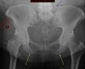
"whiskering" enthesopathy of the iliac crests (red arrows), ischial tuberosities (yellow), trochanters (black), and iliolumbar ligament (blue). The sacroiliac joints are normal.
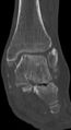
Ankle deltoid ligament ossification and tibiotalar and talocalcaneal joint arthrosis
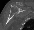
Humeral head and rotator cuff insertion hyperostosis


Extraspinal involvement can occur. It may also be possible for this to occur in the absence of spinal involvement. Practically any extraspinal articular or bony structure may be involved. When compared to osteoarthritis, DISH affects normally not seen in OA, there is marked hypertrophic change, and it affects extra-articular sites. Changes include bony proliferation (whiskering), ligamentous ossification, and periarticular osteophytosis. The peripheral enthesopathy of DISH is not normally as painful as the peripheral enthesitis of ankylosing spondyloarthritis. Clinically enthesitis can be distinguished by the presence of warm soft-tissue swelling, with an emphasis on "soft tissue" as there may be swelling related to hyperostosis.[3]
- Pelvis and hip: Iliac crest, ischial tuberosity, trochanter, iliolumbar and sacrotuberous ligaments, inferior portion of the sacroiliac joint (periarticular osteophytosis), lateral acetabulum, and superior pubic margins.[3] There may be heterotopic bone formation subsequent to total hip joint replacement.[1]
- Knees: Patella, ligamentous insertions, and the tibial tuberosity.[3]
- Ankles and feet: Dorsal surface of the talus, dorsal and medial navicular, lateral and plantar cuboid, base of the fifth metatarsal.[3]
- Shoulder: Deltoid tuberosity, inferior glenoid, inferior distal clavicle, and coracoclavicular ligament.[1]
- Elbow: Olecranon surface with normal range of motion, and medial and lateral epicondyles. [3]
- Hand: phalangeal tuft broadening and arrow heading, enlarged sesamoid bones, tubular bone cortical thickening, exostoses, joint capsule, proximal phalangeal enthesopathy.[1]
- Skull: inner table of the frontal bones called hyperostosis frontalis interna.[1]
Laboratory findings
Blood tests are usually normal or nondiagnostic. The following tests are normal or nondiagnostic: full blood count, ESR, calcium, phosphorus, growth hormone, liver function tests, parathyroid hormone, fluoride, protein eletrophoresis. HLA-B27 is not increased over the background rate.[1]
Diagnosis
The diagnosis is made radiologically. It most commonly affects the thoracic spine. There may be suggestive symptoms, and signs such as spinal stiffness and reduced range of motion. Clinical features include: palpable bony spurs (e.g. at the calcaneus, patella, or olecranon); hyperostotic olecranon spurs (at the medial and lateral epicondyles with recurrent epicondylitis); soft tissue mass that is adherent to the quadriceips, patella, or Achilles tendon; generalised peripheral entheses tenderness; recurrent achilles tendinopathy; recurrent shoulder "bursitis"; dysphagia; myelopathy in those with ossification of the posterior longitudinal ligament; stridor.[1]
The characteristic radiographic findings are flowing linear calcification and ossification along the anterior or right anterolateral aspects which continue along the disc spaces and involve at least four contiguous vertebrae. The disc height spaces should be largely preserved. Ankylosis is frequently incompletely. There can be interdigitating areas of protruding disc material in the flowing ossifications. There should be no sacroiliitis or facet joint ankylosis. There may be enthesopathy of the iliac crest, ischial tuberosities, and greater trochanters. Spur formation can occur in the appendicular skeleton including the olecranon, calcaneus, and patellar tendon. "Whiskering" enthesophytes may be seen.[1]
A lateral thoracic spine x-ray is normally the first investigation. Try and view old chest x-ray films if available, as the changes may not be reported. When cervical spine, lumbar spine, or extra-spinal disease is suspected then obtain radiographic imaging of these areas. A chest radiograph will detect 75% of cases of DISH.[1]
Classification Criteria
There are multiple classification criteriae. The most commonly used is the Resnick criteria, followed by the Utisnger criteria. Resnick criteria does not include appendicular skeleton involvement but relies exclusively on axial radiographic findings with involvement of at least four contiguous vertebrae. Utsinger only requires two contiguous vertebrae as probable DISH when in the presence of peripheral enthesopathy. For both criteria the radiographic findings have to be differentiated from other conditions, see the differential diagnosis section. See a 2017 review of the classification criteria by Kuperus et al, who highlighted the lack of consensus on the diagnosis of early DISH. [6]
| Authors | Criteria | Comment |
|---|---|---|
| Resnick [7] |
|
end stage disease, most widely used |
| Utsinger [8] |
Definite
|
lower threshold, second most widely used |
| Kuperus[9] | Uses a scoring system and algorithm using CT to classify no DISH, early DISH, and definite DISH. See Reference for free access article and movie | Validated over a five year period for picking up early disease, with a sensitivity for early DISH of 96% and specificity of 83%. |
Differential Diagnosis


The following is the complete differential diagnosis, with the diseases in bold the most important conditions to consider.[1]
- Degenerative spine disease with osteophytosis otherwise known as spondylosis deformans. This is the most common condition that should be considered. The spurs may look similar but usually the anterior longitudinal ligament is not involved in degenerative disease. In degenerative disease there may be marginal sclerosis of the vertebral body, vacuum phenomena, and disc calcification. There may be overlap.
- Ankylosing spondylitis. Misdiagnosis both ways can occur. In AS the bony bridges are normally slender and vertical ("bamboo spine"). The syndesmophytes involve the outer margin of the annulus fibrosis without affecting the anterior longitudinal ligament (marginal syndesmophytes). Vertical bone growth can also occur in DISH however, but horizontal growth (nonmarginal syndesmophytes) is more common. Sacroiliac and apophyseal joint disease may occur but this is not seen in DISH. Sacroiliac involvement in AS involves the synovial inferior two-third portion of the joint, and occurs early on. Another differentiating feature seen in DISH but not ankylosing spondylitis, is bony excrescences adjacent to peripheral joints including the knee and elbow. On MRI bone marrow oedema and fat deposition may occur in both conditions.
- Ossification of the posterior longitudinal ligament. This may occur in up to 50% of patients with DISH, and is seen in the cervical spine. It can also occur independently or in association with spondyloarthropathies. It is more common in East Asian patients. Symptoms are often neurological in nature with myelopathy and radiculopathy, and surgery is often required in these instances. Asymptomatic patients can be monitored. Thoracic and lumbar OPLL can also occur.
- Acromegaly
- Hypoparathyroidism
- Fluorosis may cause paraspinal ligament calcification.
- Ochronosis
- Retinoid arthropathy causing skeletal hyperostosis, predominantly affecting the cervical spine.
- Trauma
- X-linked hypophosphatemic osteomalacia
- Melorheostosis
| DISH | Ankylosing spondylitis | |
|---|---|---|
| Radiographic Features | ||
| Syndesmophytes (Outer annulus fibrosis ossification) | Absent* | Unusual |
| Radiographs | "Flowing candle wax" | "Bamboo spine", squaring of vertebral bodies, "shiny corners" at attachment of annulus fibrosus (Romanus lesions), "Dagger spine" (ossification of interspinal ligaments) |
| Disc space | Preservation | Cervical spine will show ossification of disc space |
| Hyperostosis | Very frequent | Frequent |
| SI joint involvement | Ligamentous obliteration | Synovial obliteration and erosions |
| ALL ossification | Very frequent | Unusual |
| PLL ossification | Very frequent | Frequent |
| Enthesopathies (whiskering) with erosions | Absent | Very frequent |
| Enthesopathies (whiskering) without erosions | Very frequent | Frequent |
| Apophyseal joint obliteration | Absent | Very frequent |
| Clinical Features | ||
| Aetiology | Idiopathic | Autoimmune |
| Age group | Middle aged >45 years | Younger patients <30 years |
| Limitation of spinal mobility and chest expansion | Frequent | Very frequent |
| Pain | Unusual | Very frequent |
| Laboratory results | Inconclusive genetic studies, non-specific results | Strong association with HLA-B27. High ESR and CRP |
| Associated conditions | Insulin resistance and diabetes | Autoimmune conditions e.g. iritis, uveitis, ulcerative colitis |
| *Some sources like orthobullets define syndesmophytes as any ligamentous ossification and state that DISH has non-marginal syndesmophytes, and AS has marginal syndesmophytes | ||
Treatment
There are no randomised controlled trials on treatment modalities in DISH. On review of the literature in August 2020 there was practically no published research that was not expert opinion on the non-surgical management of DISH. Research is hindered by the fact that the most commonly used Resnik criteria is for end stage disease. Treatment may include reassurance, physical therapy, pharmacological therapy, steroid injections,[1] as well as control of any associated metabolic disorder.
- Cervical DISH
Surgery may be required for anterior cervical osteophytes causing airway, cervical cord, or oesophageal symptoms. Ossification of the posterior longitudinal ligament may also require surgery if associated with myelopathic symptoms. Patients with cervical spine involvement should be carefully counselled as to potential concerning symptoms that may indicate myelopathy, as well as symptoms of dysphagia and stridor. If undergoing airway or gastrointestinal procedures, patients with cervical spine disease may be at higher risk of complications.[1]
- Heterotopic ossification
Patients with DISH have a propensity to develop heterotopic ossification at the site of joint arthroplasty. Some surgeons employ NSAIDs and perioperative irradiation, but this has not been systematically studied.
- Lumbar spinal stenosis plus lumbar DISH
One study showed no increased risk of surgery in the setting of lumbar spine DISH with lumbar spinal stenosis. However there was an increased risk of reoperation following successful surgery (22% vs 7.5%).[11] Another study found that lumbar DISH was an independent factor of having primary surgery.[12]
- Other considerations
Bone density is increased in DISH due to the artifact resulting from measurement in areas with substantial osteophytosis.[1]
Prognosis
The prognosis is generally good, however there is an increased rate of disability. In a large number of people it is an asymptomatic finding. Rarely potentially devastating complications can occur such as myelopathy and spinal fracture through ankylosed bone.[1]
Summary
- DISH is a progressive non inflammatory disease causing ossification of spinal ligaments and peripheral entheses, with a predilection for the thoracic spine, and takes 10 years to reach end-stage disease.
- Radiographic changes can appear with or without symptoms. Typical changes are flowing linear ossification of the anterior longitudinal ligament at the anterolateral vertebral bodies, with a right sided predominance in the thoracic spine. Ossification can also occur in other spinal ligaments. Ossification of the posterior longitudinal ligament occurs only in cervical spine disease and is seen best on CT. Peripheral ossification can occur at entheseal locations causing whiskering changes.
- Restriction of movement and pain are both common, but the disease may be asymptomatic. In some severe cases the disability incurred can be similar to severe ankylosing spondylitis.[Level 3]
- The aetiology is unknown, but there is some association with insulin resistance and metabolic derangement. There appear to be genetic, metabolic, mechanical and vascular factors, as well as changes in signalling pathways.[Level 3]
- The prevalence varies between populations and the setting of research, but it is a disease of middle age. Rates of DISH are very high amongst Pacific Islanders and Maori.[Level 3]
- Red flag symptoms as a sign of cervical spine disease are dysphagia, odynophagia, dysphonia, and stridor. In addition, ossification of the posterior longitudinal ligament can occur causing myelopathic symptoms.
- Careful consideration should be made when considering the differential diagnosis, with the main differentials being degenerative changes and ankylosing spondylitis, as misdiagnosis is common between these three conditions in all directions. There is no consensus on the diagnostic criteria. [Level 5]
- DISH may be treated with reassurance, physical therapy, pharmacological therapy, steroid injections, and control of any associated metabolic disorder. Research on prevention and treatment is practically non-existent. [Level 5]
Quiz
References
- ↑ 1.00 1.01 1.02 1.03 1.04 1.05 1.06 1.07 1.08 1.09 1.10 1.11 1.12 1.13 1.14 1.15 1.16 1.17 1.18 1.19 1.20 1.21 1.22 1.23 1.24 1.25 1.26 1.27 1.28 1.29 Simon M Helfgott et al. Diffuse idiopathic skeletal hyperostosis (DISH). In: UpToDate, Post, TW (Ed), UpToDate, Waltham, MA, March 2020.
- ↑ Bateman et al.. Diffuse idiopathic skeletal hyperostosis (DISH): Increased prevalence in Pacific Islanders. Journal of medical imaging and radiation oncology 2018. 62:188-193. PMID: 29024571. DOI.
- ↑ 3.0 3.1 3.2 3.3 3.4 3.5 3.6 3.7 3.8 3.9 Olivieri et al.. Diffuse idiopathic skeletal hyperostosis: differentiation from ankylosing spondylitis. Current rheumatology reports 2009. 11:321-8. PMID: 19772826. DOI.
- ↑ Olivieri et al.. Diffuse idiopathic skeletal hyperostosis may give the typical postural abnormalities of advanced ankylosing spondylitis. Rheumatology (Oxford, England) 2007. 46:1709-11. PMID: 17938137. DOI.
- ↑ Yaniv et al.. The natural course of bridging osteophyte formation in diffuse idiopathic skeletal hyperostosis: retrospective analysis of consecutive CT examinations over 10 years. Rheumatology (Oxford, England) 2014. 53:1951-7. PMID: 24158753. DOI.
- ↑ Kuperus et al.. Classification criteria for diffuse idiopathic skeletal hyperostosis: a lack of consensus. Rheumatology (Oxford, England) 2017. 56:1123-1134. PMID: 28371859. DOI.
- ↑ Resnick & Niwayama. Radiographic and pathologic features of spinal involvement in diffuse idiopathic skeletal hyperostosis (DISH). Radiology 1976. 119:559-68. PMID: 935390. DOI.
- ↑ Utsinger et al.. Diffuse skeletal abnormalities in Forestier disease. Archives of internal medicine 1976. 136:763-8. PMID: 938166.
- ↑ Kuperus et al.. Criteria for Early-Phase Diffuse Idiopathic Skeletal Hyperostosis: Development and Validation. Radiology 2019. 291:420-426. PMID: 30938626. DOI. Full Text.
- ↑ Colin Woon et al. DISH (Diffuse Idiopathic Skeletal Hyperostosis). Accessed 19/8/20. Available from: https://www.orthobullets.com/spine/2045/dish-diffuse-idiopathic-skeletal-hyperostosis
- ↑ Yamada et al.. Diffuse Idiopathic Skeletal Hyperostosis Extended to the Lumbar Segment Is a Risk Factor of Reoperation in Patients Treated Surgically for Lumbar Stenosis. Spine 2018. 43:1446-1453. PMID: 29481381. DOI.
- ↑ Yamada et al.. Diffuse idiopathic skeletal hyperostosis is associated with lumbar spinal stenosis requiring surgery. Journal of bone and mineral metabolism 2019. 37:118-124. PMID: 29327302. DOI.
Literature Review
 Literature Review
- Reviews from the last 7 years: review articles, free review articles, systematic reviews, meta-analyses, NCBI Bookshelf
- Articles from all years: PubMed search, Google Scholar search.
- TRIP Database: clinical publications about evidence-based medicine.
- Other Wikis: Radiopaedia, Wikipedia Search, Wikipedia I Feel Lucky, Orthobullets,

