Enthesis
The enthesis (plural entheses) is the interface between tendon/ligament and bone. See also about the myotendinous junction, the connection between muscle and tendon
Structure and Function

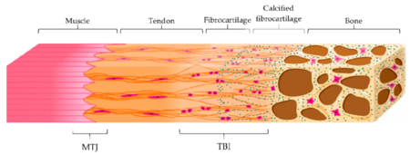
The musculoskeletal system as a whole has both hard and soft tissues. The interfaces between these tissues have gradients in order to reduce stress concentrations at the junction sites. The interfaces between tendons/ligaments and bone are called entheses, while the interfaces between tendons and muscles are called myotendinous junctions (MTJ).
The structure of the enthesis varies depending on the tissue present at the skeletal attachment site. There are two main categories of entheses: fibrous entheses, and the fibrocartilaginous entheses. Most entheses in the body are fibrocartilaginous.
The function of the enthesis is to dissipate stress away from the interface.
Fibrous Enthesis
The fibrous enthesis is where there is direct attachment to either bone or periosteum via dense fibrous fibrous connective tissue. This tissue is similar in composition to the tendon midsubstance.
It consists of tendons and ligaments being connected through acute angles to bones with collagen fibres extending directly from the periosteum, termed Sharpey's fibres.
This type of enthesis is commonly found in tendons that are attached to the metaphyses diaphyses of long bones. The insertion site is typically over large surface areas.
Examples are the deltoid attachment to the humerus, adductor magnus insertion to the linear aspera of the femur, and the pronator teres.
Fibrocartilaginous Enthesis

In this type of enthesis, there is a layer of fibrocartilage acting as a transition from the fibrous tendon to bone. The tendon is directly inserted into the bone and the tendon tissue close to the bone calcifies.
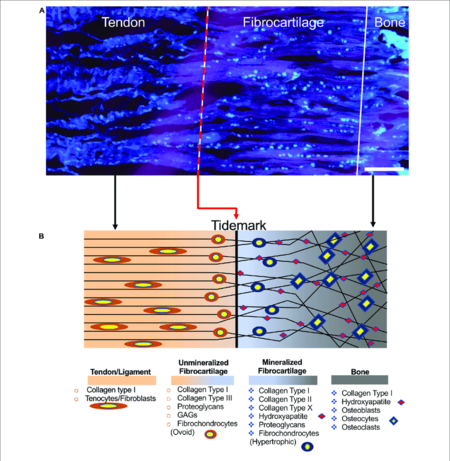
The fibrocartilaginous enthesis consists of a progressive mineralisation gradient that is organised into four zones.
- Tendon/ligament: This region consists of pure dense fibrous connective tissue. There are longitudinally oriented fibroblasts and a parallel arrangement of collagen fibres (type I and III). There is also elastin and proteoglycans within the ground substance.
- Unmineralised fibrocartilage (also called uncalcified fibrocartilage): contains various collagens (types I, II, III) and proteoglycans (mostly aggrecans with associated chondroitin 4- and 6- glycosaminoglycans). The collagen fibres become increasingly randomly arranged. Fibroblasts and tenocytes are replaced by ovoid-shaped aligned fibrochondrocytes. It acts as a shock absorber to dissipate the stresses generated by the tendon. It is avascular.
- Mineralised fibrocartilage. There are hypertrophic chondrocytes surrounded by predominantly type II but also types I and X collagens and aggrecans. It is avascular and irregular with interlocking with bone. It provides the mechanical integrity of the insertion to provide for a transition of mechanical force across the insertion. It also acts as a barrier against blood vessels in the bone, and prevents communication between osteocytes and tendon cells.
- Bone. This consists of osteocytes, osteoblasts, and osteoclasts, in a mineralised matrix.
The boundary between the unmineralised and mineralised fibrocartilage zones is called the tidemark where there is an abrupt transition between soft and hard tissues. The thickness of the junction is around 500 µm.
The fibrocartilaginous type of enthesis is more common, and is also prone to overuse injuries. It is found in the epiphyses and apophyses. Examples are the rotator cuff and achilles tendons. It is not that fibrocartilaginous attachments as a design entity are more likely to lead to injury compared to fibrous attachments, but that insertion sites that are at risk for injury have fibrocartilaginous attachments in order to reduce the risk of injury.
Tendons that insert onto the epiphyses and apophyses tend to have more variable ranges of insertion angles during their movement. This creates increased stress with bending of the collagen fibres. The fibrocartilage acts to dissipate this stress. In other words it is a structural adaptation. Compare for example the fibrous insertion of the deltoid muscle which doesn't change its angle greatly with abduction, to the fibrocartilaginous insertion of the supraspinatus which does change its angle greatly with abduction.
Clinical Applications
A disease process affecting the entheses is called enthesopathy or enthesitis. Enthesopathy is also associated with spondyloarthropathies. Many commonly seen tendinopathies are strictly enthesopathies.
Common locations for injury are the rotator cuff, the anterior cruciate ligament, the Achilles tendon, the medial collateral ligament of the knee, tennis elbow, and jumper's knee. The gradations present in fibrocartilaginous entheses are not regenerated during tendon to bone healing. Instead, fibrovascular scar tissue forms, composed primarily of type III collagen, which is mechanically weaker and more prone to failure than the original enthesis.
The optimal treatment strategies are as yet unknown.
Further Reading
See Also
References
- ↑ Bianchi E, Ruggeri M, Rossi S, Vigani B, Miele D, Bonferoni MC, Sandri G, Ferrari F. Innovative Strategies in Tendon Tissue Engineering. Pharmaceutics. 2021 Jan 11;13(1):89. doi: 10.3390/pharmaceutics13010089. PMID: 33440840; PMCID: PMC7827834.
- ↑ Sensini, Alberto et al. “Tissue Engineering for the Insertions of Tendons and Ligaments: An Overview of Electrospun Biomaterials and Structures.” Frontiers in bioengineering and biotechnology vol. 9 645544. 2 Mar. 2021, doi:10.3389/fbioe.2021.645544
Literature Review
- Reviews from the last 7 years: review articles, free review articles, systematic reviews, meta-analyses, NCBI Bookshelf
- Articles from all years: PubMed search, Google Scholar search.
- TRIP Database: clinical publications about evidence-based medicine.
- Other Wikis: Radiopaedia, Wikipedia Search, Wikipedia I Feel Lucky, Orthobullets,


