File:LFCN ultrasound.jpg: Difference between revisions
From WikiMSK
No higher resolution available.
LFCN_ultrasound.jpg (600 × 452 pixels, file size: 71 KB, MIME type: image/jpeg)
No edit summary |
No edit summary |
||
| (One intermediate revision by the same user not shown) | |||
| Line 1: | Line 1: | ||
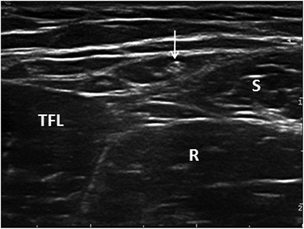
Transverse ultrasound image of the LFCN lying within the intermuscular space between the tensor fasciae latae muscle and the sartorius.<br><small>LFCN: lateral femoral cutaneous nerve; TFL: tensor fasciae latae muscle; S: Sartorius; R: rectus femoris.</small><ref>{{#pmid:23171132}}</ref> | |||
Latest revision as of 19:22, 8 April 2021
Transverse ultrasound image of the LFCN lying within the intermuscular space between the tensor fasciae latae muscle and the sartorius.
LFCN: lateral femoral cutaneous nerve; TFL: tensor fasciae latae muscle; S: Sartorius; R: rectus femoris.[1]
File history
Click on a date/time to view the file as it appeared at that time.
| Date/Time | Thumbnail | Dimensions | User | Comment | |
|---|---|---|---|---|---|
| current | 18:30, 8 April 2021 | | 600 × 452 (71 KB) | Jeremy (talk | contribs) | File uploaded with MsUpload |
You cannot overwrite this file.
File usage
The following page uses this file:

