Finger Anatomy: Difference between revisions
(Created page with "{{Partial}} ==Dorsal Aspect== {{multiple image | align = right | image1 = Finger extensor mechanism dorsal view.jpg | width1 = 200 | alt1 = | caption1 = Dorsal view | image2...") |
No edit summary |
||
| Line 1: | Line 1: | ||
{{Partial}} | {{Partial}} | ||
==Dorsal Aspect== | ==Dorsal Aspect== | ||
{{multiple image | {{multiple image | ||
| Line 51: | Line 50: | ||
{{Article derivation|article-link=https://www.e-ultrasonography.org/journal/view.php?doi=10.14366/usg.15051|article=Current status of ultrasonography of the finger|author=Seun Ah Lee et al|license-link=https://creativecommons.org/licenses/by-nc/3.0/|license=CC-BY-NC}} | {{Article derivation|article-link=https://www.e-ultrasonography.org/journal/view.php?doi=10.14366/usg.15051|article=Current status of ultrasonography of the finger|author=Seun Ah Lee et al|license-link=https://creativecommons.org/licenses/by-nc/3.0/|license=CC-BY-NC}} | ||
[[Category:Hand and Wrist Anatomy]] | [[Category:Hand and Wrist Anatomy]] | ||
[[Category:Upper Limb Anatomy]] | |||
Latest revision as of 17:20, 30 April 2022
Dorsal Aspect
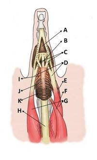
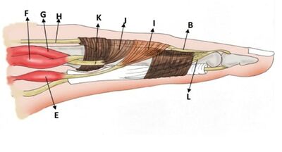
The extensor hood stabilises the extensor tendon at the MCPJ, whereas the dorsal extensor apparatus stabilises the PIPJ.
Extensor Hood
The extensor hood (expansion) is a triangular aponeurosis on the dorsal aspect of the proximal phalanx by which the extensor tendons insert onto the phalanges.
The extensor hood surrounds the distal metacarpal head and proximal phalanx and serves to hold the extensor tendons in place and allow the extensors, lumbricals, and interossei to effect extension at the proximal and distal interphalangeal joints.
The extensor hood is made up of the sagittal bands, oblique fibres, and transverse fibres. The sagittal bands are the main stabilisers of the extensor hoot and maintain the extensor tendons in the midline of the metacarpal head during flexion and extension of the fingers. The ulnar and radial sagittal bands exert tensile forces in opposite directions during flexion, which keeps the extensor tendon in apposition with the metacarpal bone.[2]
Muscles
The two main components of the extensor tendons are extrinsic muscles and intrinsic muscles. The extrinsic muscles, which originate from the forearm and elbow and insert into the hand, comprise the extensor digitorum communis, extensor indicis proprius, and extensor digiti quinti minimi. The intrinsic muscles, which originate and insert into the hand, include the interosseous and lumbrical muscles.[2]
Slips
The long extensor tendons of the posterior forearm (extensor digitorum, extensor indicis, and extensor digiti minimi) cross the MCPJ with their deepest fibres adhering partially to the posterior joint capsule and the remaining tendon bulk passing the joint. Distal to the MCP joint, the extensor tendons flatten and fan out as they traverse the dorsal proximal phalanx and separate into three slips.
- One central slip: inserts on to the base of the middle phalanx
- Two lateral slips: diverge laterally around the central slip to insert on the base of the distal phalanx
The extrinsic extensor tendon continues in the central and lateral slips, the while the intrinsic extensor tendons contribute to the formation of the lateral slips. After the lateral slips conjoin with the intrinsic muscles, they are termed conjoint tendons and form the terminal tendon that inserts into the dorsal aspect of the distal phalangeal base.
On the extensor surface of the thumb, there is no extensor expansion proper: the tendons of the extensor pollicis brevis and longus muscles are inserted separately in the proximal and distal phalanx respectively. However, the tendon of the extensor pollicis longus muscle does receive a fibrous expansion from the abductor pollicis brevis and adductor pollicis muscles which serves a similar purpose as the digital extensor expansion: to hold the extensor tendon in place on the dorsal phalangeal surface.[3]
Volar Aspect
Two flexor tendons are present on the volar side of the fingers: the flexor digitorum profundus (FDP) and the flexor digitorum superficialis (FDS). They run within the fibrous tendon sheath. Once the FDS enters the tendon sheath, it is separated into two tendons that surround the FDP and insert into the shaft of the middle phalanx. The FDP is initially located deep in the FDS, and then enters the cleft formed by the division of the FDS and inserts into the distal phalangeal base.
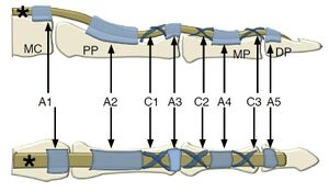
The flexor tendon sheath extends from the metacarpal neck to the DIP. A series of retinacular structures in which the sheath is thickened at five specific points, form the annular pulley system (pulleys A1-A5), while another three form the cruciate pulley system (pulleys C1-C3). These pulleys prevent excursion of the flexor tendons from the MCP and IP joints during finger flexion. The A2 pulley is the thickest, strongest, and longest annular pulley.
The MCP, PIP, and DIP joints are reinforced by the volar plate, which is composed of thick, resistant fibrocartilage. The volar plates limit finger hyperextension.
All of the digital articulations have one essential feature in common: They are designed to function in flexion. Each joint has firm collateral ligaments bilaterally and a thick anterior capsule reinforced by a fibrocartilaginous structure known as the palmar (volar) plate. By comparison, the dorsal capsule is thin and lax.
Lateral Aspect
The main stabilizers of the MCP and PIP joints include the volar plate and collateral ligaments. The collateral ligaments of the MCP joint are taut during flexion and lax during extension, allowing abduction and adduction. The collateral ligament of the PIP joints consists of the proper and the accessory collateral ligaments; the proper collateral ligament is taut during flexion, whereas the accessory collateral ligament is taut during extension.
The MCP joint of the thumb is stabilized by the volar plate, collateral ligaments (similar to the PIP and MCP joints of other fingers), and additional musculotendinous structures. The adductor pollicis has a strong tendinous point of insertion into the PP and the volar plate/sesamoid complex. Some of its fibres also contribute to the adductor aponeurosis, which covers the ulnar collateral ligament (UCL) of the MCP joint of the thumb.
See Also
References
Part or all of this article or section is derived from Current status of ultrasonography of the finger by Seun Ah Lee et al, used under CC-BY-NC
- ↑ Ditsios K, Konstantinou P, Pinto I, Karavelis A, Kostretzis L (2017) Extensor Mechanism’s Anatomy at the Metacarpophalangeal Joint. MOJ Orthop Rheumatol 8(4): 00319. DOI: 10.15406/mojor.2017.08.00319
- ↑ 2.0 2.1 Lee SA, Kim BH, Kim SJ, Kim JN, Park SY, Choi K. Current status of ultrasonography of the finger. Ultrasonography. 2016 Apr;35(2):110-23. doi: 10.14366/usg.15051. Epub 2015 Nov 24. PMID: 26753604; PMCID: PMC4825212.
- ↑ A, A., Bell, D. Extensor expansion. Reference article, Radiopaedia.org. (accessed on 06 Feb 2022) https://doi.org/10.53347/rID-70476


