Hip Joint
| |
| Hip Joint | |
|---|---|
| Synonym | Acetabulofemoral joint |
| Primary Type | Synovial Joint |
| Secondary Type | Ball and Socket Joint |
| Bones | Ilium, Femur |
| Ligaments | Ischiofemoral, iliofemoral, pubofemoral, transverse acetabular, and ligamentum teres |
| Muscles | 27 musculotendinous units are involved in hip function |
| Innervation | Femoral, obturator and superior gluteal nerves, and nerve to quadratus femoris |
| Vasculature | Branches of the medial and lateral circumflex femoral, superior and inferior gluteal arteries and obturator arteries |
| ROM | Flexion 140°, extension 20°, Internal rotation 30°, external rotation 40°, abduction 50°, adduction 30° |
| Volume | 1-10mL |
| Conditions | Hip Osteoarthritis, Hip Labral Tear, Gluteal Tendinopathy |
The hip joint is a synovial joint between the femoral head and the acetabulum of the pelvis.
Bones and Articulations
The hip is a ball and socket joint. The ball is the spherical head of the femur, while the socket is the concave acetabulum. The cartilage of the hip is viscoelastic which means that the amount of deformation that occurs to the cartilage is inversely proportional to the magnitude of load and rate that the load is applied (e.g. high load leads to little deformation).
Acetabulum
The acetabulum is horseshoe shaped with the acetabular notch in the inferior region. The acetabular cartilage is thicker in the periphery where it merges with the labrum. It is particularly thick in the superior-anterior region of the dome. Articulation with the femoral head only occurs on the horseshoe-shaped hyaline cartilage.
The acetabulum is angled obliquely forward: it is directed anterior, lateral, and inferior. A mal-aligned acetabulum doesn't adequately cover the femoral head and this can lead to chronic dislocation and early onset osteoarthritis.
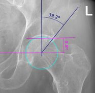
Lateral Centre-Edge: How much coverage the acetabulum provides over the femoral head in the frontal/coronal plane is measured by various parameters on radiographs such as the centre edge angle which normally measures 20-45° (Figure 1). A normal centre edge angle provides a protective shelf over the femoral head in the coronal plane, while a small angle means that there is less coverage leading to risk of dislocation and injury. A larger angle such as in pincer femoro-acetabular impingement protects against developing hip osteoarthritis.
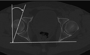
Acetabular Version: In the horizontal/axial plane femoral head coverage can be measured by angle of acetabular anteversion and this measures 20° on average (Figure 2). An increase in this angle such as in hip dysplasia is associated with decreased joint stability and an increased risk of dislocation, labral injury, and chondral injury. Version can also be seen on frontal views, with the crossover sign being consistent with acetabular retroversion, and the absence of the crossover sig being consistent with acetabular anteversion. Acetabular version can vary greatly with pelvic tilt. (See Hip Radiograph)
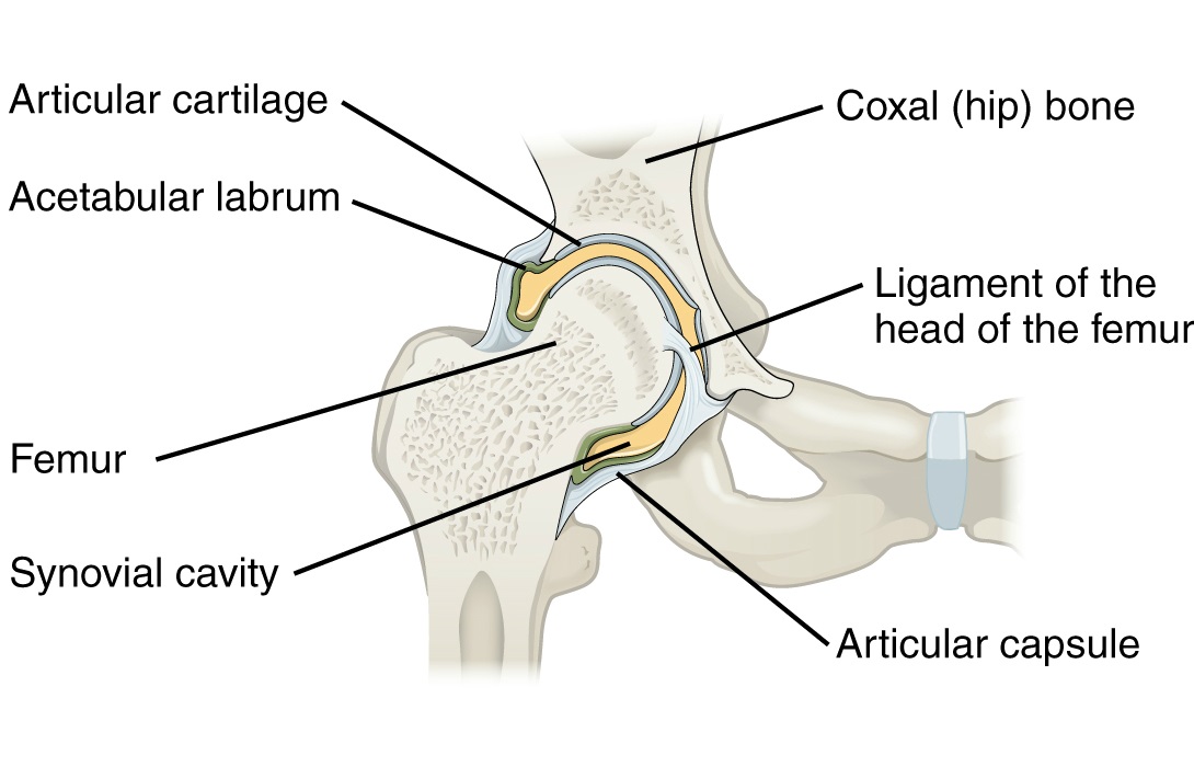
Labrum: The labrum is a fibrocartilaginous lip that encircles and deepens the acetabulum. It blends with the transverse acetabular ligament which spans the acetabular notch. It helps to contain the femoral head in place in extremes of motion, especially in flexion. Along with the capsule, the labrum acts as a load-bearing structure during flexion. The labrum helps to maintain a negative pressure vacuum within the joint that is produced by the acetabular fossa which helps to stabilise the joint. Individuals with a deficient labrum may have instability and capsular laxity.
Femoral Head and Neck
The femur is the largest and strongest bone in the body.
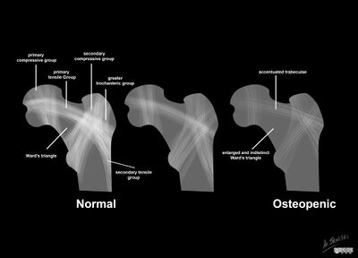
The head and neck are composed of cancellous bone. The trabecular patterns of growth follow the course of stress lines along the bone with maximum trabeculae developing along lines of maximum stress (both compressive and tensile). There are five types of trabeculae - two compressive, two tensile, and the greater trochanteric trabeculae.
The two compressive systems are the principal medial and secondary lateral trabecular systems. The joint reaction force is parallel with the medial compressive system. The epiphysial plates are at right angles to the medial system. The role of the lateral compressive system is to resist the compressive force on the femoral head that is produced by contraction of the hip abductors.
There are also trabecular lines corresponding to tensile forces. The primary tensile system forms an arc extending from the lateral margin of the greater trochanter to the inferior aspect below the fovea. There is a central area bounded by three systems called Ward's triangle which is a zone of weakness in the anatomical neck.
The cortical bone is thin on the superior surface, and progressively thickens towards the inferior region. During the aging process the cortical bone becomes thinner and cancellated while the trabeculae are gradually resorbed.
Femoral Head: The femoral head is the convex part of the ball-and-socket joint of the hip. It forms two-thirds of a sphere. The head is covered by cartilage which is thickest on the medial-central aspect surrounding the fovea (the ligamentum teres attaches to the fovea), and is thinnest towards the periphery. With smaller loads the load-bearing area is concentrated at the periphery; while with larger loads the load-bearing area is concentrated at the centre and anterior and posterior horns. (due to the viscoelastic properties of the cartilage). Hip dysplasia leads to improper load distribution which leads to osteoarthritis and further changes in load distribution.
Femoral Neck: The femoral neck is the narrowest part and internally it is primarily composed of trabecular bone, and so it is the weakest component of the femur. The femur angles medially downward to aid in single-leg support during walking and running.
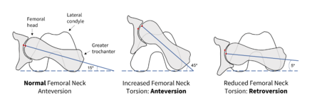
The proper functioning of the hip is partly determined by the angle of the femoral neck to the femoral shaft. This can be demonstrated by the neck-to-shaft angle otherwise known as the angle of inclination in the frontal/coronal plane (figure 4), and the torsion angle in the horizontal/axial plane (figure 5).
The Angle of Inclination: The angle of inclination is a frontal/coronal plane measurement and is normally 90-135° but is greater at birth and in childhood. An increased angle is called coxa valga, and a decreased angle is called coxa vara. For both of these variations there is a change in the biomechanics of the hip related to changes in the moment arm used by the hip abductors and changes in shear forces (See Nordin and Frankel).
Femoral Version: Femoral version is the angle of the femoral neck in the axial plane relative to the angle of the knee (through the femoral condyles) in the horizontal/axial plane, which can be measured on CT. The normal angle is 10-20° of femoral anteversion. Newborns have even more anteversion, and this is a reflection of the medial rotatory migration of the lower limb bug that occurs in embryological development. With excessive femoral neck anteversion, a portion of the femoral head is uncovered and there is a tendency for the leg to internally rotate during gait to keep the femoral head in the acetabular cavity (in-toeing). With retroversion, there is a tendency for the leg to externally rotate during gait (out-toeing). These are both quite common in childhood but individuals usually outgrow it.
See youtube video on femoral torsion.
Hip Capsule and Ligaments
The labrum is a ring of fibrocartilage that contributes to joint stability. Compared to the glenoid fossa of the shoulder, the acetabular fossa is much deeper and so is more stable.
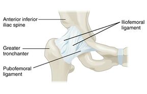
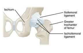
The hip capsule is composed of three capsular ligaments: two anterior called the iliofemoral and pubofemoral ligaments, and one posterior called the ischiofemoral ligament. The hip capsule is an important stabiliser of the hip, especially at extremes of motion to prevent dislocation. These major ligaments act to twist the head of the femur into the acetabulum during hip extension.
The capsule is thickened anterosuperiorly which is the site of predominant stress. It is relative thin and loosely attached posteroinferiorly. The ligaments are twisted around the neck in a clockwise direction. This reflects embryological development. This means that the ligaments are tightest in medial rotation and extension, and loosest in flexion and lateral rotation
There is also the transverse acetabular ligament which prevents inferior dislocation of the femoral head.
Within the joint capsule is the ligamentum teres which attaches directly from the rim of the acetabulum to the head of the femur.
Bursae
The iliopsoas bursa and deep trochanteric bursa are the most prominent bursae. The iliopsoas is located between the iliopsoas and articular capsule and reduces friction between these two structures. The deep trochanteric bursa reduces friction between the greater trochanter of the femur and the gluteus maximus.
Movements
The pelvic girdle is analogous to the shoulder girdle in that it positions the hip for effective movement. The shoulder girdle is made up of multiple different joints, while the pelvis is non-jointed. Nevertheless, the pelvis is able to rotate in all three planes. With pelvic rotation, the acetabular position is changed so that it is directed towards the impending femoral movement. Posterior pelvic tilt enables increases hip flexion by placing the head of the femur in front of the pelvis. Anterior pelvic tilt increases hip extension, and lateral pelvic tilt increases lateral movements. The pelvis also moves in coordination with the spine.
There are a variety of two-joint muscles. Two-joint muscles function most effectively at one joint when the position of the other joint stretches the muscle slightly.
- Flexion: iliacus, psoas major, pectineus, rectus femoris, sartorius, and tensor fascia lata.
- Iliopsoas (iliacus and psoas major) is the major hip flexor.
- Rectus femoris is a two-joint muscle and is active during both hip flexion and knee extension; it works better as a hip flexor with the knee is flexion.
- Sartorius is also a two-joint muscle.
- Extension: Gluteus maximus, biceps femoris, semitendinosus, and semimembranosus (latter three are the hamstrings).
- Gluteus maximus is active only with hip flexion
- Hamstrings are two-joint muscles, and aid in hip extension and knee flexion.
- Abduction: Gluteus medius and minimus
- Gluteus medius: major abductor
- Gluteus minimus: assists.
- Both muscles stabilise the pelvis during the support phase of walking and running and with standing on one leg.
- Adduction:Adductor longus, adductor brevis, adductor magnus, gracilis
- Active during swing phase of gait to bring the foot below the body's centre of gravity before the support phase. Also active during stair and hill climbing.
- Gracilis: strap muscle contributes to knee flexion.
- Adductor longus, adductor brevis, adductor magnus: also contributes to flexion and lateral hip rotation especially with a medially rotated femur.
- Lateral rotators: piriformis, gemellus superior, gemellus inferior, obturator internus, obturator externus, quadratus femoris
- Stronger than medial rotators.
- Medial rotators: gluteus minimus, tensor fascia lata, semitendinosus, semimembranosus, gluteus medius
- Horizontal abduction
- Horizontal abduction
