Hip Labral Tear: Difference between revisions
No edit summary |
No edit summary |
||
| Line 19: | Line 19: | ||
Labral tears are present in 22% of athletes with groin pain and 55% of those with mechanical symptoms.<ref name="brukner"/> [[Ligamentum Teres Tear]]s are found in 70% of sportspeople's hips when undergoing arthroscopy for FAI or labral tears.<ref name=brukner/> | Labral tears are present in 22% of athletes with groin pain and 55% of those with mechanical symptoms.<ref name="brukner"/> [[Ligamentum Teres Tear]]s are found in 70% of sportspeople's hips when undergoing arthroscopy for FAI or labral tears.<ref name=brukner/> | ||
Whilst common in people with hip pain, labral tears are also common in individuals who may be asymptomatic. Therefore, the difficulty is in deciding which labral tears are clinically relevant. In a study on asymptomatic hockey players, 56% had labral tears<ref>{{ | Whilst common in people with hip pain, labral tears are also common in individuals who may be asymptomatic. Therefore, the difficulty is in deciding which labral tears are clinically relevant. In a study on asymptomatic hockey players, 56% had labral tears,<ref>{{#pmid:18641370}}</ref> and a study on 70 asymptomatic young health professionals found labral tears in 38.6%.<ref>{{Cite journal|last=Lee AJJ|first=|last2=Armour P|last3=Thind D|last4=Coates M|last5=Kang ACL|date=2015|title=The prevalence of acetabular labral tears and associated pathology in a young asymptomatic population|url=|journal=Bone Jt J.|volume=97-B(5)|pages=623-627|doi=10.1302/0301-620X.97B5.35166|pmid=|via=|doi-access=}}</ref> | ||
A study of young asymptomatic volunteers in a ski town demonstrated labral tears in approximately 69%. Other hip pathology was also common; 24% had a chondral lesion. In addition, 95% of those with FAI also had a labral tear.<ref name="register" /> | A study of young asymptomatic volunteers in a ski town demonstrated labral tears in approximately 69%. Other hip pathology was also common; 24% had a chondral lesion. In addition, 95% of those with FAI also had a labral tear.<ref name="register" /> | ||
| Line 62: | Line 62: | ||
Magnetic resonance arthrography (MRA) is the most accurate imaging modality, but the gold standard remains the arthroscopic exam<ref name="brukner" />. MRA has a sensitivity and accuracy of 90% and 91%, versus 30% and 36% with conventional MRI.<ref>{{#pmid:11479747}}</ref> Findings are best assessed in the presence of a joint effusion or contrast, as fluid can be seen penetrating through the labral contour or chondrolabral junction. Other findings may be present such as a displaced labrum, chondral injury, paralabral cyst, or stripping of the capsule. | Magnetic resonance arthrography (MRA) is the most accurate imaging modality, but the gold standard remains the arthroscopic exam<ref name="brukner" />. MRA has a sensitivity and accuracy of 90% and 91%, versus 30% and 36% with conventional MRI.<ref>{{#pmid:11479747}}</ref> Findings are best assessed in the presence of a joint effusion or contrast, as fluid can be seen penetrating through the labral contour or chondrolabral junction. Other findings may be present such as a displaced labrum, chondral injury, paralabral cyst, or stripping of the capsule. | ||
The newer 3.0 Tesla MRI systems have a 93-95% sensitivity and 65-89% specificity for diagnosing labral tears which are confirmed by arthroscopy, and are about as accurate as 1.5T MRA.<ref>{{Cite journal|last=Chopra A|first=|last2=Grainger AJ | The newer 3.0 Tesla MRI systems have a 93-95% sensitivity and 65-89% specificity for diagnosing labral tears which are confirmed by arthroscopy, and are about as accurate as 1.5T MRA.<ref>{{Cite journal|last=Chopra A|first=|last2=Grainger AJ|date=2018|title=Comparative reliability and diagnostic performance of conventional 3T magnetic resonance imaging and 1.5T magnetic resonance arthrography for the evaluation of internal derangement of the hip|url=|journal=Eur Radiol.|volume=28(3)|pages=963-971|doi=10.1007/s00330-017-5069-4|pmid=|via=|doi-access=}}</ref><ref>{{Cite journal|last=Wen C; Huishu Y|first=|date=2019|title=3.0T MR arthrography in diagnosis of acetabular labral tears confirmed by arthroscopy|url=https://epos.myesr.org/poster/esr/ecr2019/C-1651|journal=EPOS European Society of Radiology|volume=|pages=|doi=|pmid=|via=|doi-access=}}</ref> | ||
More recently, some authors have proposed that hip traction ultrasonography is a useful adjunct to MRA for diagnosis of labral tears.<ref>{{Cite journal|last=Billham J; Cornelson SM; Koch A; Nunez M; Estrada P; Kettner N|first=|date=2020|title=Diagnosing acetabular labral tears with hip traction sonography: a case series|url=|journal=J Ultrasound|volume=|pages=|doi=10.1007/s40477-020-00446-x|pmid=|via=|doi-access=}}</ref> The literature for this requires further development. | More recently, some authors have proposed that hip traction ultrasonography is a useful adjunct to MRA for diagnosis of labral tears.<ref>{{Cite journal|last=Billham J; Cornelson SM; Koch A; Nunez M; Estrada P; Kettner N|first=|date=2020|title=Diagnosing acetabular labral tears with hip traction sonography: a case series|url=|journal=J Ultrasound|volume=|pages=|doi=10.1007/s40477-020-00446-x|pmid=|via=|doi-access=}}</ref> The literature for this requires further development. | ||
An intraarticular injection of local anaesthetic with or without a glucocorticoid can aid in the diagnostic process if pain is ablated following injection. If an MRA is performed then anaesthetic can be injected along with the contrast and a pain diary can be ascertained. This technique helps to assist with determining whether a labral tear is symptomatic. A study on 75 patients who had MRA + diagnostic local anaesthetic block showed a 80% sensitivity and 83% specificity for labral tear as diagnosed by arthroscopy.<ref>{{Cite journal|last=Kheterpal AB|first=|last2=Bunnell KM|last3=Husseini JS | An intraarticular injection of local anaesthetic with or without a glucocorticoid can aid in the diagnostic process if pain is ablated following injection. If an MRA is performed then anaesthetic can be injected along with the contrast and a pain diary can be ascertained. This technique helps to assist with determining whether a labral tear is symptomatic. A study on 75 patients who had MRA + diagnostic local anaesthetic block showed a 80% sensitivity and 83% specificity for labral tear as diagnosed by arthroscopy.<ref>{{Cite journal|last=Kheterpal AB|first=|last2=Bunnell KM|last3=Husseini JS|date=2020|title=Value of response to anesthetic injection during hip MR arthrography to differentiate between intra- and extra-articular pathology|url=|journal=Skeletal Radiol.|volume=49(4)|pages=555-561|doi=10.1007/s00256-019-03323-9|pmid=|via=|doi-access=}}</ref> The test is most useful if it is positive with a PPV of 91%. | ||
==Management== | ==Management== | ||
Revision as of 05:32, 22 June 2021
Acetabular labral tears are a common cause of hip and groin pain in athletes and in those with degenerative hip joint conditions. Optimal diagnosis and management remains unknown, and due to the very high prevalence of asymptomatic tears the clinician is urged to treat the patient not the MRI.
Anatomy
- Main article: Acetabular Labrum Anatomy

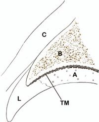
The acetabular labrum is a fibrocartilaginous structure that seals the central hip joint from the periphery, keeps the synovial fluid within the central compartment, and creates a negative pressure within the joint. It increases the depth, volume, surface area, and congruity of the hip joint. The negative pressure helps to resist subluxation of the femoral head and increases stability.
Any disruption of the labrum can negatively affect articular cartilage health and joint stability.[2] On the other hand, labral tears are extremely common in the asymptomatic population, and it is still not completely known which types of tears are clinically important and may become symptomatic.[3]
The vascular supply originates from the radial branches of a periacetabular periosteal vascular ring, which arises mainly from the superior and inferior gluteal arteries. The vessels course over the periosteum, and penetrate the through the capsule and connective tissue, ending at the free edge of the labrum. This means that the vascular supply remains intact even with a tear at the chondrolabral junction.[4]
Labral tears start at the junction between the labrum and cartilage, termed the watershed region. The vascular supply enters through the synovium at the point of reflection of the capsule onto the peripheral border of the labrum. The vessels only penetrate the outermost layer of the labrum. [5]
Nociceptive fibres are found at the highest density in the anterior labrum, but course mostly anterosuperiorly to posterosuperiorly. This is also the typical area of labral pathology. The free nerve endings are located primarily at the labral base, and decrease peripherally, being mostly superficial on the chondral surface. The fibres arise mainly from the obturator nerve and the nerve to the quadratus femoris. Proprioceptive end organs are also found within the labrum.[4]
Epidemiology
Labral tears are present in 22% of athletes with groin pain and 55% of those with mechanical symptoms.[6] Ligamentum Teres Tears are found in 70% of sportspeople's hips when undergoing arthroscopy for FAI or labral tears.[6]
Whilst common in people with hip pain, labral tears are also common in individuals who may be asymptomatic. Therefore, the difficulty is in deciding which labral tears are clinically relevant. In a study on asymptomatic hockey players, 56% had labral tears,[7] and a study on 70 asymptomatic young health professionals found labral tears in 38.6%.[8]
A study of young asymptomatic volunteers in a ski town demonstrated labral tears in approximately 69%. Other hip pathology was also common; 24% had a chondral lesion. In addition, 95% of those with FAI also had a labral tear.[3]
Pathogenesis
There are two general mechanisms of injury to the acetabular labrum.[2]
- Traumatic tears: A single event of significant trauma. This normally involves forced resistance of hip flexion while kicking or running (for example in Rugby).
- Degenerative tears: Repetititve injury and microtrauma in an osteoarthritic, dysplastic hip or in a hip with FAI.
Pathology normally occurs in the weightbearing anterosuperior aspect of the labrum.[2] There are several thoughts as to the reasons for this prediliction. There is reduced thickness of the anterior labrum. Femoroacetabular impingement normally causes anterior impingement. Repetitive twisting and pivoting is a factor. Owing to anteversion of the acetabulum, there is reduced bony support anteriorly which may also increase the shear forces on the labrum. Increased forces are also placed on the anterior labrum during the final stages of the stance phase of gait and in more than 5 degrees of hip extension.[6]
Healing of the labrum has been demonstrated in animal studies.[9]
Classification
Tears occur at the articular labral junction. However there there is a classification system for tears that categorises them based on aetiology, morphology, location, and histology.
- Aetiology: traumatic, degenerative, idiopathic, congenital, or dysplastic
- Morphology: radial flap, radial fibrillated, longitudinal, peripheral, or unstable. The two radial tears are most common involving the free margins of the labrum. Unstable tears cause mechanical symptoms.
- Location: anterior, posterior, or superior, or by clock face
- Histology: Type I tears are a detachment of the labrum from the acetabular rim cartilage, perpendicular to the articular surface at the transition zone. Type II is a cleavage tear or tears within the labrum substance, extending perpendicular to the surface of the labrum.[5]
The most common injury in the western world is the watershed lesion. This is an anterior labral tear present with anterior acetabular chondral injury, seen after minor trauma. Posterior labrum tears are normally associated with a discrete episode of trauma.[5]
It is still not known which types of tears are pathological and which may be normal variants.[3] The tear location in respect to vascular supply is important when considering healing potential.[6] However the location of the tear has not been proven to have an effect on prognosis.[5]
Clinical Features
The most common symptom is groin pain that is exacerbated by athletic activity. Pain is normally located in the anterior hip or groin, and is often described as sharp. Uncommonly it can can cause mechanical symptoms such as catching, clicking, locking, [2] or giving way.[5] Some patients may describe buttock pain, which is more consistent with a posterior labral tear[5] Pain can occur in particular with activities involving aggressive hip flexion such as jumping or sprinting. Pain can sometimes occur when fatigued such as during a long distance run, or running up hill. Some patients may report groin pain with sitting, transitioning between standing from sitting, or when descending stairs. Some activities of daily living may be affected such as putting on shoes or stockings while sitting. [2]
Neumann et al in 2007 evaluated 100 adults with mechanical hip symptoms using MRA. Symptoms were groin pain (48%), groin and buttock pain (7%), clicking (23%), catching (56%), stiffness (5%), snapping (12%), popping (13%), cracking (2%), and giving way (7%). Labral tears were found in 66% of these patients.[10]
Examination has poor sensitivity and sensitivity.[6] Features are pain with hip flexion and anterior impingement tests. No test is specific for labral injury, and signs and symptoms overlap with FAI. Some useful tests are repeated hip flexion, hip flexion against resistance, FABER, and FADDIR testing. Reduced internal rotation may suggest hip joint osteoarthritis.[2][6]
The ligamentum teres test can be performed if a tear of this structure is suspected. This involves the patient being supine, flexing the hip to full flexion minus 30 degrees, abducting to full abduction minus 30 degrees, them moving the hip internally and externally through full range of motion. A positive test is reported pain.[6]
Imaging
Plain X-ray is unable to demonstrate labral pathology but is useful for determining whether there may be predisposing factors that contribute to acetabular labral tears such as FAI or DDH. X-rays should include standing anteroposterior, cross-table lateral or Dunn lateral, and a false profile view.
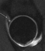
Magnetic resonance arthrography (MRA) is the most accurate imaging modality, but the gold standard remains the arthroscopic exam[6]. MRA has a sensitivity and accuracy of 90% and 91%, versus 30% and 36% with conventional MRI.[11] Findings are best assessed in the presence of a joint effusion or contrast, as fluid can be seen penetrating through the labral contour or chondrolabral junction. Other findings may be present such as a displaced labrum, chondral injury, paralabral cyst, or stripping of the capsule.
The newer 3.0 Tesla MRI systems have a 93-95% sensitivity and 65-89% specificity for diagnosing labral tears which are confirmed by arthroscopy, and are about as accurate as 1.5T MRA.[12][13]
More recently, some authors have proposed that hip traction ultrasonography is a useful adjunct to MRA for diagnosis of labral tears.[14] The literature for this requires further development.
An intraarticular injection of local anaesthetic with or without a glucocorticoid can aid in the diagnostic process if pain is ablated following injection. If an MRA is performed then anaesthetic can be injected along with the contrast and a pain diary can be ascertained. This technique helps to assist with determining whether a labral tear is symptomatic. A study on 75 patients who had MRA + diagnostic local anaesthetic block showed a 80% sensitivity and 83% specificity for labral tear as diagnosed by arthroscopy.[15] The test is most useful if it is positive with a PPV of 91%.
Management
Conservative Management
An initial trial of non-operative management is recommended.[6] Physical therapy is aimed as minimising dynamic FAIT and reducing abnormal loads on the labrum. [2]
- Pelvic tilt should be optimised to reduce dynamic FAI. This can be done through pelvic girdle strengthening, gluteus maximus activation, abductor control, transversus abdominis and rectus abdominis activation, iliopsoas stretching, adductor stretching, rectus femoris stretching, core control, and pelvic floor control. [4] Exercises should start unloaded and progress with more load added.[6]
- Optimise sagittal balance with the spine and pelvis, factors remote from the hip joint affecting biomechanics.[4]
- Gait retraining can also be considered in order to reduce excessive hip extension at the end of the stance phase of gait.[6]
Activity modification should also be discussed. The patient should be advised to avoid repetitive hip flexion, adduction, abduction, and rotation at end range.
The evidence for physical therapy in symptomatic hip labral tears is very limited. A physical therapy progression program was described in a case series of four young patients with positive outcomes. Phase 1 emphasised pain control, education in trunk stabilization, and correction of abnormal joint movement. Phase 2 focused on muscular strengthening, recovery of normal range of motion (ROM), and initiation of sensory motor training. Phase 3 emphasised advanced sensory motor training, with sport-specific functional progression.[16]
PRP
A small prospective study of 8 people reported positive outcomes with the injection of PRP at the site of labral tear at 8 weeks of follow up.[17]
Surgery
Arthroscopic surgery can be considered upon failure of conservative management and is aimed at treating the labrum and the underlying cause.[4] In athletes with FAI, surgery can be considered early if their sport requires a range of motion not achieved before impingement symptoms occur.[6] Surgical options include debridement, repair, and reconstruction with an allograft or autograft (normally from the ITB or hamstring tendons). Where possible the labrum should be restored rather than simply debrided in order to restore the normal hip joint seal and this probably provides better outcomes. The removal of damaged tissue and replacement with a graft may aid in pain reduction due to the presence of nociceptive fibres within the labrum. Proprioceptive fibres are also removed and so it is important to be mindful of overly stressing the graft postoperatively. The labrum can be repaired in certain instances. [4]
There are no randomised sham controlled trials for arthroscopy surgery, and so surgical treatment remains unproven. A systematic review and meta-analysis of 8 level 3-4 observational studies of labral reconstruction showed positive outcomes, and no difference between allograft vs autograft.[18]
Contraindications are contraindications to hip preservation surgery rather than labral repair itself. They include significant hip osteoarthritis, or in combination with uncorrected dysplasia. There is no role for prophylactic repair in the asymptomatic population.[4]
Bottom Line
- Physical therapy is the first line treatment.[Level 5]
- Surgical repair or grafting is second line, avoiding simple debridement if possible. [Level 3]
References
- ↑ Su et al.. Diagnosis and treatment of labral tear. Chinese medical journal 2019. 132:211-219. PMID: 30614856. DOI. Full Text.
- ↑ 2.0 2.1 2.2 2.3 2.4 2.5 2.6 Johnson, R. Approach to hip and groin pain in the athlete and active adult. In: UpToDate, Post, TW (Ed), UpToDate, Waltham, MA, 2020.
- ↑ 3.0 3.1 3.2 Register et al.. Prevalence of abnormal hip findings in asymptomatic participants: a prospective, blinded study. The American journal of sports medicine 2012. 40:2720-4. PMID: 23104610. DOI.
- ↑ 4.0 4.1 4.2 4.3 4.4 4.5 4.6 Harris. Hip labral repair: options and outcomes. Current reviews in musculoskeletal medicine 2016. 9:361-367. PMID: 27581790. DOI. Full Text.
- ↑ 5.0 5.1 5.2 5.3 5.4 5.5 Groh & Herrera. A comprehensive review of hip labral tears. Current reviews in musculoskeletal medicine 2009. 2:105-17. PMID: 19468871. DOI. Full Text.
- ↑ 6.00 6.01 6.02 6.03 6.04 6.05 6.06 6.07 6.08 6.09 6.10 6.11 Brukner. Clinical Sports Medicine. 4th Edition. McGraw-Hill. 2012
- ↑ Feeley et al.. Hip injuries and labral tears in the national football league. The American journal of sports medicine 2008. 36:2187-95. PMID: 18641370. DOI.
- ↑ Lee AJJ; Armour P; Thind D; Coates M; Kang ACL (2015). "The prevalence of acetabular labral tears and associated pathology in a young asymptomatic population". Bone Jt J. 97-B(5): 623–627. doi:10.1302/0301-620X.97B5.35166.
- ↑ Miozzari et al.. Effects of removal of the acetabular labrum in a sheep hip model. Osteoarthritis and cartilage 2004. 12:419-30. PMID: 15094141. DOI.
- ↑ Neumann et al.. Prevalence of labral tears and cartilage loss in patients with mechanical symptoms of the hip: evaluation using MR arthrography. Osteoarthritis and cartilage 2007. 15:909-17. PMID: 17383908. DOI.
- ↑ Petersilge. MR arthrography for evaluation of the acetabular labrum. Skeletal radiology 2001. 30:423-30. PMID: 11479747. DOI.
- ↑ Chopra A; Grainger AJ (2018). "Comparative reliability and diagnostic performance of conventional 3T magnetic resonance imaging and 1.5T magnetic resonance arthrography for the evaluation of internal derangement of the hip". Eur Radiol. 28(3): 963–971. doi:10.1007/s00330-017-5069-4.
- ↑ Wen C; Huishu Y (2019). "3.0T MR arthrography in diagnosis of acetabular labral tears confirmed by arthroscopy". EPOS European Society of Radiology.
- ↑ Billham J; Cornelson SM; Koch A; Nunez M; Estrada P; Kettner N (2020). "Diagnosing acetabular labral tears with hip traction sonography: a case series". J Ultrasound. doi:10.1007/s40477-020-00446-x.CS1 maint: multiple names: authors list (link)
- ↑ Kheterpal AB; Bunnell KM; Husseini JS (2020). "Value of response to anesthetic injection during hip MR arthrography to differentiate between intra- and extra-articular pathology". Skeletal Radiol. 49(4): 555–561. doi:10.1007/s00256-019-03323-9.
- ↑ Yazbek et al.. Nonsurgical treatment of acetabular labrum tears: a case series. The Journal of orthopaedic and sports physical therapy 2011. 41:346-53. PMID: 21335929. DOI.
- ↑ De Luigi et al.. Use of Platelet-Rich Plasma for the Treatment of Acetabular Labral Tear of the Hip: A Pilot Study. American journal of physical medicine & rehabilitation 2019. 98:1010-1017. PMID: 31162277. DOI.
- ↑ Rahl et al.. Outcomes After Arthroscopic Hip Labral Reconstruction: A Systematic Review and Meta-analysis. The American journal of sports medicine 2020. 48:1748-1755. PMID: 31634004. DOI.
Literature Review
- Reviews from the last 7 years: review articles, free review articles, systematic reviews, meta-analyses, NCBI Bookshelf
- Articles from all years: PubMed search, Google Scholar search.
- TRIP Database: clinical publications about evidence-based medicine.
- Other Wikis: Radiopaedia, Wikipedia Search, Wikipedia I Feel Lucky, Orthobullets,


