Internal Disc Disruption
Internal disc disruption is a condition that affects lumbar intervertebral discs. There are isolated, radial fissures that penetrate from the nucleus pulposus through to the anulus fibrosus, however the outer anulus is not breached.
It is a separate concept to disc degeneration. While there are many similar processes and features, internal disc disruption is a response to injury that occurs either through single or repetitive compressive loads.
Aetiology
End-plate fracture
Internal disc disruption occurs following injury to the overlying vertebral end plate. This can occur following a single sudden compressive force, or through fatigue failure from repetitive lower forces. End plate fracture can occur with as few as 100 repetitions with compressive loads as low as 50% to 60% of the ultimate tensile strength of the end plate.
Once end-plate fracture has occurred, disc degradation can occur. While the exact sequence of events is uncertain, it is well established that end-plate fracture is an important inciting event. Mechanistic theories include inflammatory response, and a reduction in the pH leading to an increased activity of metalloproteases.
Following matrix degradation, it is thought that the matrix isn't able to brace the anulus from buckling inwards. It may be that continued normal activities of every day life progressively leads to tearing of the anulus in this unsupported state.
Biomechanics
Biomechanically there are significant changes in the internal stresses. In the normal state, while the outer portion of the anulus has no compression stresses, the inner portion of the anulus and the nucleus pulposus have uniform stresses, with a small peak in the posterior anulus.
In the setting of internal disc disruption, the disc is not able to bear compression loads normally. There are irregular stresses that have reduced amplitudes, even reaching zero in some areas. This pattern is seen immediately following end-plate fracture.
Correlation with Pain
Radial fissures aren't degenerative or age related changes as they are independent of these two factors. They are strongly associated with the disc being painful on provocative discography.
Nociception is thought to occur through interaction of nuclear material with the outer third of the annulus, which is where nociceptive fibres are located, which is why grade I and II fissures are less likely to be painful. Both chemical and mechanical nociception is theorised to occur.
Classification

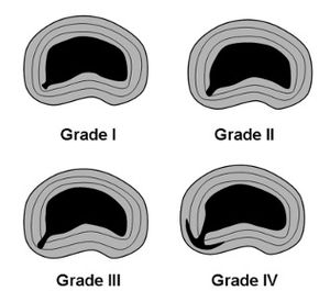
Fissures can be graded based on the extent of penetration into the anulus.
- Grade I: reaches the inner third of the anulus
- Grade II: reaches the middle third of the anulus
- Grade III: reaches the outer third of the anulus
- Grade IV: the fissure spreads circumferentially around the annulus
Grade III and IV fissures are most likely to be painful.
Epidemiology
Internal disc disruption is common in those with chronic low back pain who have invasive tests, with different studies reporting prevalences of 39%, 26%, and 42%.
Imaging
The gold standard for the diagnosis of internal disc disruption in through post-discography CT scan imaging. With discography, contrast is injected into the nucleus. With the contrast in place, CT imaging shows up any fissures and their grade. post-discography CT is not commonly done in the clinical setting.
MRI can be used to look for two specific findings: modic changes and high intensity zones. Both of these features are independently correlated with pain, however they are not exclusive to the symptomatic population. They are not pathognomic for that segment being the cause of pain, but are highly specific.
- Modic changes occur around the endplates and represent current (modic type I) or past (modic type II) inflammatory changes.
- High intensity zones represent circumferential annular tears.
Bibliography
- Bogduk N. Degenerative joint disease of the spine. Radiol Clin North Am. 2012 Jul;50(4):613-28. doi: 10.1016/j.rcl.2012.04.012. PMID: 22643388.
- Bogduk N, Aprill C, Derby R. Lumbar discogenic pain: state-of-the-art review. Pain Med. 2013 Jun;14(6):813-36. doi: 10.1111/pme.12082. Epub 2013 Apr 8. PMID: 23566298.
- ↑ Bogduk N. Degenerative joint disease of the spine. Radiol Clin North Am. 2012 Jul;50(4):613-28. doi: 10.1016/j.rcl.2012.04.012. PMID: 22643388.

