Lateral Femoral Cutaneous Nerve
| |
| Lateral Femoral Cutaneous Nerve | |
|---|---|
| Nerve Type | Sensory nerve |
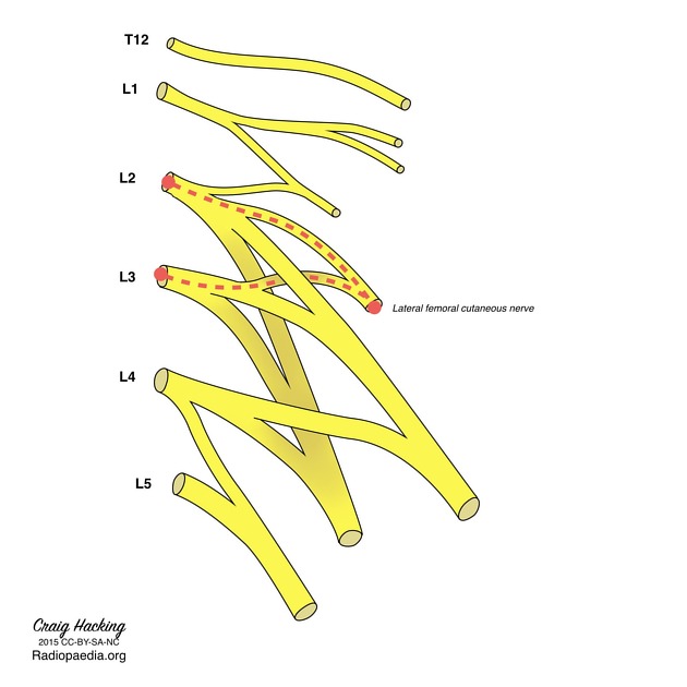
| Origin | Posterior divisions of L2 and L3 spinal nerves |
| Course | 3cm above inguinal ligament, to within fibrous tissue of iliac fascia, to below or through the inguinal ligament, to the thigh just medial to the ASIS, to over the sartorius deep to the fascia lata. |
| Major Branches | Anterior and posterior branches |
| Sensory innervation | Anterolateral thigh |
| Conditions | Lateral Femoral Cutaneous Nerve Entrapment |
The lateral femoral cutaneous nerve of the thigh (LFCN) is a pure sensory nerve that supplies the anterolateral thigh. The topics of Meralgia Paraesthetica and lateral femoral cutaneous nerve injections are dealt with elsewhere.
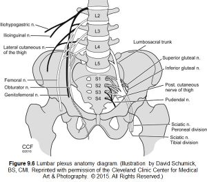
Origin and Course
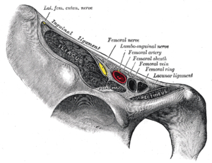
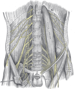
The LFCN arises from the lumbar plexus, originating from the posterior divisions of the L2 and L3 spinal nerves (the anterior divisions contribute to the obturator nerve). It starts from between the superficial and deep parts of the psoas muscle and goes around the pelvis on the iliacus muscle between two layers of fascia. It then travels through an “aponeuroticofascial tunnel” that courses from the iliopubic tract to the inguinal ligament, under the inguinal ligament or through an opening in the lateral aspect of the ligament, medial to the ASIS. Around 10cm below the inguinal ligament, it exits through the superficial fascia of the thigh, passing over the sartorius deep to the fascia lata, and divides into anterior and posterior branches. It terminates in the skin of the anterolateral thigh.
Sensory Distribution
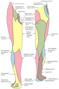
The LFCN has two branches, the anterior (larger) branch, and the posterior (smaller) branch, supplying the anterior and posterior aspects of the lateral thigh as far inferiorly as the knee.
The anterior branch typically contains all the L3 fibres of the nerve and supplies the anterolateral thigh to the knee. It has terminal twigs that also donate to the patellar plexus
The posterior branch typically contains all L2 fibres and continues down the posterolateral aspect of the thigh along the iliotibial tract contributing terminal filaments that pass across the lateral and posterior surfaces of the thigh
Motor Distribution
There are no motor fibres.
Anatomic Variability
The LFCN has marked anatomical variability. It is fused with the genitofemoral nerve in around 2% of cases which predisposes to increased vulnerability to lumbar sympathetic block. It has intra-abdominal branching with one vertebral origin, and one distributing branch at the level of the inguinal ligament in 86% of cases. It has a variable exit out of the abdomen. 4% of the time it is posterior to the ASIS across the iliac crest, 27% is medial to the ASIS superficial to the origin of the sartorius, 23% it is medial to the ASIS within the sartorius muscle, 26% it is medial to the origin of the sartorius muscle deep to the inguinal ligament, and 20% it is very medial deep to the inguinal ligament but superficial to the iliopsoas fascia. The distance from the ASIS at the level of the inguinal ligament varies between 2.3cm lateral, to 6.2cm medial, with an average distance of 1.4 ± 1.5 cm. There can be as many as five inguinal branches, in 28% it branches before the inguinal ligament. The angle between the pelvic and femoral portions is 100 ± 10°. In its relationship to the sartorius, it exits through the muscle 22% of the time, or exits at its tendon of origin in 23%. With respect to the superficial thigh fascia, 88% of the time the LFCN is deep to it below the ASIS, 3% are superficial to it, and 9% are not found. There are anterior and posterior branches 54% of the time, no posterior branch 36% of the time. The area of innervated skin is highly variable. There is side to side symmetry in 65%.
Other Relevant Structures
- Transversus abdominis muscle
- This may partially originally from the iliacus fascia, and so abdominal contraction can increase stress on the LFCN.
- Iliopubic tract
- The LFCN can be compressed here, which is analogous to median nerve entrapment in carpal tunnel syndrome.
- Inguinal ligament
- Many structures converse on the inferior border of the oblique aponeurosis. This includes the fascia lata and also the origin of sartorius.
- Muscular compartment of the inguinal region
- Between the inguinal ligament, the iliopectineal arch, and the ilium, through which the LFCN, femoral nerve, and iliopsoas muscle reach the leg
- Deep circumflex vessels
- The LFCN passes deep to these vessels as it approaches the ASIS.
References
- Trescot, Andrea. Peripheral nerve entrapments : clinical diagnosis and management. Switzerland: Springer, 2016.
- Dr Michael Stewart and Dr Chamath Ariyasinghe et al. Lateral femoral cutaneous nerve. Radiopaedia.org
