Lateral Hip Rotators: Difference between revisions
From WikiMSK
No edit summary |
No edit summary |
||
| Line 1: | Line 1: | ||
[[File:Posterior Hip Muscles.png|thumb|right|Posterior hip muscles]] | |||
There is surprisingly little consensus on the anatomy of the lateral hip rotators{{#pmid:26032283|yoo}} | There is surprisingly little consensus on the anatomy of the lateral hip rotators{{#pmid:26032283|yoo}} | ||
| Line 17: | Line 18: | ||
|- | |- | ||
|} | |} | ||
==Lumbosacral Plexus Images== | ==Lumbosacral Plexus Images== | ||
Revision as of 19:42, 12 August 2020

There is surprisingly little consensus on the anatomy of the lateral hip rotators[1]
| Muscle | Origin | Insertion | Innervation |
|---|---|---|---|
| Piriformis | Anterior surface of sacrum between and laterally to the anterior sacral foramina | Superior boundary of greater trochanter | Nerve to the piriformis (S1-S2) |
| Gemellus Superior | Ischial spine | Upper edge of Obturator internus muscle tendon (indirectly greater trochanter) | Nerve to obturator internus (L5-S2) |
| Internal Obturator | Medial surface of obturator membrane and the surrounding bone | Medial surface of greater trochanter | Nerve to obturator internus (L5-S2) |
| Gemellus Inferior | Just above the tuberosity of the ischium | Lower edge of Obturator internus muscle tendon (indirectly greater trochanter) | Nerve to quadratus femoris (L4-S1) |
| Quadratus Femoris | Lateral edge of the tuberosity of the ischium | Intertrochanteric crest | Nerve to quadratus femoris (L4-S1) |
| External Obturator | Lateral surface of obturator membrane and the ischiopubic ramus | Trochanteric fossa | Posterior branch of obturator nerve (L3-L4) |
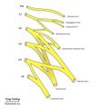
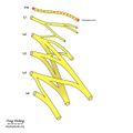
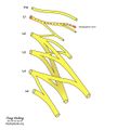
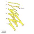
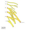
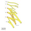
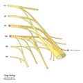
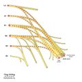
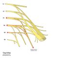
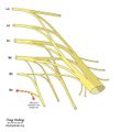
Lumbosacral Plexus Images
Lumbar Plexus
Lumbar Plexus
Subcostal nerve
Iliohypogastric nerve
Ilioinguinal nerve
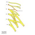
Genitofemoral nerve
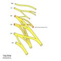
Lateral femoral cutaneous nerve
Obturator nerve
Femoral nerve
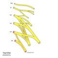
Lumbosacral trunk


Sacral Plexus
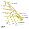
Sacral Plexus
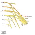
Superior gluteal nerve
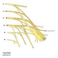
Inferior gluteal nerve
Nerve to piriformis
Sciatic nerve
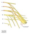
Nerve to quadratus and inferior glemellus
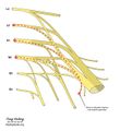
Nerve to obturator internus and superior gemellus
Posterior femoral cutaneous nerve
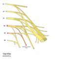
Perforating cutaneous nerve
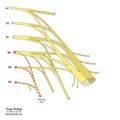
Pudendal nerve
Nerves to perineum and levator ani


