◔
Lateral Hip Rotators: Difference between revisions
From WikiMSK
No edit summary |
No edit summary |
||
| Line 32: | Line 32: | ||
{{Muscles of the Hip and Lower Limb}} | {{Muscles of the Hip and Lower Limb}} | ||
[[Category:Lower Limb | [[Category:Muscles of the Lower Limb]] | ||
Revision as of 20:20, 15 July 2021
This article is a stub.
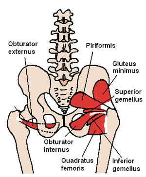
There is surprisingly little consensus on the anatomy of the lateral hip rotators[1]
| Muscle | Origin | Insertion | Innervation |
|---|---|---|---|
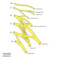
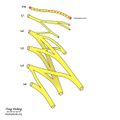
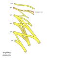
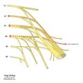
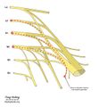
| Piriformis | Anterior surface of sacrum between and laterally to the anterior sacral foramina | Superior boundary of greater trochanter | Nerve to the piriformis (S1-S2) |
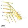
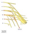
| Gemellus Superior | Ischial spine | Upper edge of Obturator internus muscle tendon (indirectly greater trochanter) | Nerve to obturator internus (L5-S2) |
| Internal Obturator | Medial surface of obturator membrane and the surrounding bone | Medial surface of greater trochanter | Nerve to obturator internus (L5-S2) |
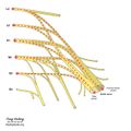
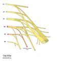
| Gemellus Inferior | Just above the tuberosity of the ischium | Lower edge of Obturator internus muscle tendon (indirectly greater trochanter) | Nerve to quadratus femoris (L4-S1) |
| Quadratus Femoris | Lateral edge of the tuberosity of the ischium | Intertrochanteric crest | Nerve to quadratus femoris (L4-S1) |
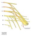
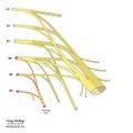
| External Obturator | Lateral surface of obturator membrane and the ischiopubic ramus | Trochanteric fossa | Posterior branch of obturator nerve (L3-L4) |
