Lumbar Instability
The term instability as it pertains to the lumbar spine is controversial. Instability is a biomechanical term.
Biomechanics
Instability is when a small load causing an abnormally large or catastrophic displacement. There is no gold standard definition for instability of the lumbar spine. The concept has been viewed through different lenses
Decreased Stiffness: (relates to terminal instability) This definition views instability as "a loss of spinal motion segment stiffness such that a force application to the structure produces a greater displacement(s) than would be seen in a normal structure, resulting in a painful condition, the potential for progressive deformity, and that places neurologic structures at risk".
This doesn't cover the idea that a small load causes a large displacement, and loss of stiffness may simply be hypermobile segments not at risk of catastrophic collapse. Bogduk suggests the term hypermobility or segmental looseness be applied here instead.
Increased Neutral Zone: (relates to instability in a normal range) "An increased neutral zone is a significant decrease in the capacity of the stabilising system of the spine to maintain the intervertebral neutral zones within the physiological limits so that there is no neurological dysfunction, no major deformity, and no incapacitating pain. "
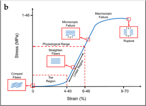
The neutral zone is similar to the length of the toe phase of the stress strain curve (see Lumbar Spine Biomechanics), with an increased neutral zone corresponding to an increased length of the toe phase. The joints are loose in early range with excessive displacement, but their ultimate strength may be normal.

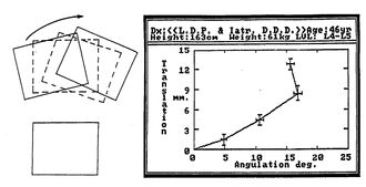
Instability Factor: (relates to instability in a normal range) Terminal instability is the behaviour of a system at its endpoint with a sense of impending failure. However the quality of movement can also be assessed during normal range not only terminal range. This can be viewed by looking at the amount of translation relative to rotation that occurs during sagittal movement of the lumbar spine. Normal lumbar segments have a uniform ratio of translation to rotation.
Instability could be seen as an abnormal change of this ratio at any point in movement within normal range, but it may be stable at end range. This type of instability can be viewed by taking serial radiographs during range of motion, and calculating the ratio of the sums of incremental movement. Mean and standard deviations have been reported. An abnormal instability factor can be defined as being outside two standard deviations. Patients with degenerative disc disease have larger instability factors compared to controls, with increased translation relative to rotation with movement from extension to flexion.[1]
This technique is very time consuming and it not used in routine clinical practice.
Anatomy
Passive stability is provided by the facet joints and ligaments, with the facet joints being the major stabilising elements in flexion. The back muscles act to increase stability by providing a compressive load which makes it more difficult for the joints to move. The multifidus has the strongest influence.
In extreme scenarios overt failures or impending failure can be detected radiographically for example in fracture-dislocation. In this situation it is readily apparent that there is disruption of the stabilising elements. However it is uncertain how to determine the threshold for instability in less extreme situations.
Force, Displacement, and Acceleration
There are both displacing and restraining forces. With lumbar movement there is a force that causes displacement. Stabilising forces act against displacement to prevent uncontrolled acceleration. Motion occurs with an appropriate combination of displacing and restraining forces, with displacement occurring over time (i.e. velocity). Velocity increases with initial movement and decreases at the end of movement eventually stopping at end range.
With reduced stiffness, the restraining forces are reduced, but the displacing forces are unchanged, and so the acceleration and velocity of movement is increased. Instability occurs if the balance between the displacing and restraining forces do not prevent inordinate displacement or the threat of failure. This type of instability occurs throughout range of motion and at end range.
An increased neutral zone occurs when there is a loss of restraints in early range but not in end range. There is a normal early velocity, but it eventually has a higher velocity with the altered ratio of displacing and restraining forces. This type of instability occurs because the terminal velocity is excessive and unexpected. This unfamiliar acceleration is alarming to the individual, even if inappropriately alarming.
When there is a loss of restraining forces at mid or late range, initial movements are normal, but there is acceleration in late rate. This late acceleration may also feel alarming.
This force-displacement model looks at acceleration being the factor that corresponds to the degree of instability, with a mismatch between proprioceptive feedback and the motor programme for movement. A particular individual may develop an increase in acceleration, but may inappropriately continue to use the previous patterns of motor activity, which are now insufficient to maintain stability. The segment now feels like it is giving way. The motor system may react crudely with a sudden jerk or catch, or the muscles may be persistently active to guard against any movement that risks accelerating the segment.
Relationship to Pain
It is uncertain how instability may relate to pain. Certainly at early range for a hypermobile segment, the segment shouldn't be painful. Pain may occur at end range when the restraining tissues are being strained. Sudden jerking pain at end range could arise if the terminal restraints are rapidly engaged rather than in a normal gradual manner, with a sudden compressive load being applied due to reactive muscle contraction. If the loss of stiffness has an underlying cause, i.e. some kind of injury, then the pain may arise from that injury, not the instability. In this scenario the pain is aggravate with movement because the injured structure is being irritated not because of instability.
Clinical vs Biomechanical Instability
The above descriptions are for biomechanical instability. Clinical instability is not equivalent to biomechanical instability. There are two meanings of clinical instability
- Relates to the patients symptoms, rather than what is biomechanically happening.
- When the biomechanical instability is clinically relevant and produces symptoms, however it doesn't imply that the instability is clinically evident.
The diagnosis of instability is dependent on biomechanical tests.
Diagnosis
A common abuse of the term instability is labelling people who have pain with movement as having instability. This is flawed because conditions can occur that are aggravated by movement but don't involve any instability. The range of motion may actually be restricted rather than excessive. There are in fact no valid criteria for diagnosing subtle forms of instability. Clinical criteria need to be compared against a criterion standard to know if they are valid. However the only criterion standard is radiographic changes and these have difficulties.
| Category | Causes | Validity |
|---|---|---|
| I | Fractures and fracture-discloations | Valid |
| II | Infection of the anterior elements | Valid |
| III | Neoplasms | Valid |
| IV | Spondylolisthesis | Controversial |
| V | Degenerative | Complicated |
Spondylolisthesis is controversial as a valid entity of instability. Patients with grade 1 and grade 2 spondylolisthesis have reduced range of motion rather than instability. Some patients have anterior translation with standing from lying, but it isn't clear if this is abnormal. Research using implanted tantalum balls for accurate landmark identification haven't found evidence of instability.
The degenerative category is the most complicated and controversial of all categories. It remains uncertain whether degenerative conditions are associated with instability and whether this can actually be diagnosed. The various sub-types of degenerative instability include rotational and retrolisthetic instability for which there are no operational criteria; and translational instability for which there are criteria. Other subtypes are post-disc excision, post-laminectomy, and post-fusion which may be more readily diagnosed. Scoliotic instability is simply a type of rotational and translational instability.
For translational instability the main problem is that there is no agreed upper limit of normal translation. 3mm, 4mm, and 5mm have been proposed. but these findings can still occur in normal individuals. The translation should be dynamic. This means that it is apparent in full flexion but not in extension, or vice versa.
A study by Kasai and colleagues tested a clinical sign called the Passive Lumbar Extension Test in 122 patients with lumbar spinal stenosis (mixed central and lateral), lumbar spondylolisthesis, and lumbar degenerative scoliosis. Immediately there are issues as to what the criterion standard are which they recognise. They diagnosed instability if the patient had one of the following, using the highest reported cut off values they could find: angular motion of 20 degrees comparing flexion to extension, translational motion of 5mm, or intervertebral end-plate angle on flexion film of -5 degrees. Patients who had none of these were classified as not having instability. The positive LR was 8.84, and negative LR was 0.17.[2]
References
Bogduk. Instability In: Clinical and radiological anatomy of the lumbar spine. Elsevier 2012
- ↑ 1.0 1.1 Weiler PJ, King GJ, Gertzbein SD. Analysis of sagittal plane instability of the lumbar spine in vivo. Spine (Phila Pa 1976). 1990 Dec;15(12):1300-6. doi: 10.1097/00007632-199012000-00012. PMID: 2149207.
- ↑ Kasai Y, Morishita K, Kawakita E, Kondo T, Uchida A. A new evaluation method for lumbar spinal instability: passive lumbar extension test. Phys Ther. 2006 Dec;86(12):1661-7. doi: 10.2522/ptj.20050281. Epub 2006 Oct 10. PMID: 17033040.


