Lumbar Spine MRI: Difference between revisions
No edit summary |
No edit summary |
||
| (14 intermediate revisions by the same user not shown) | |||
| Line 1: | Line 1: | ||
{{ | {{partial}} | ||
==Interpretation== | ==Anatomical Structures== | ||
===Sagittal Scans=== | |||
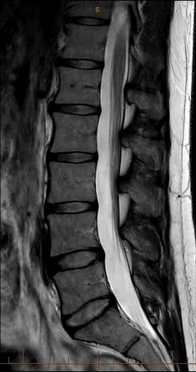
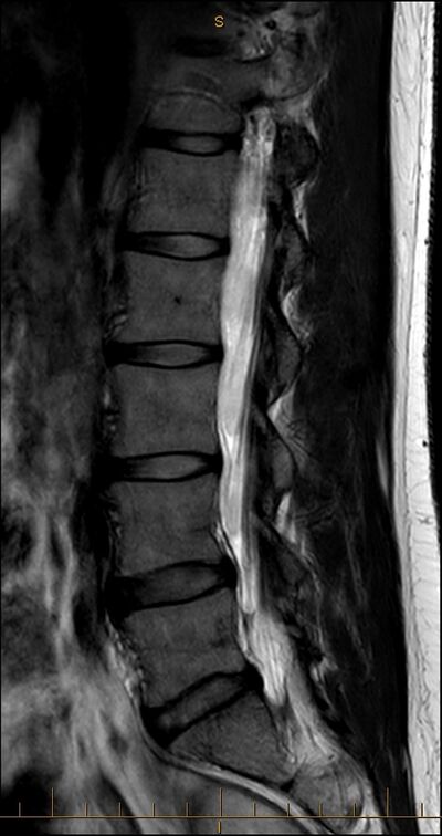
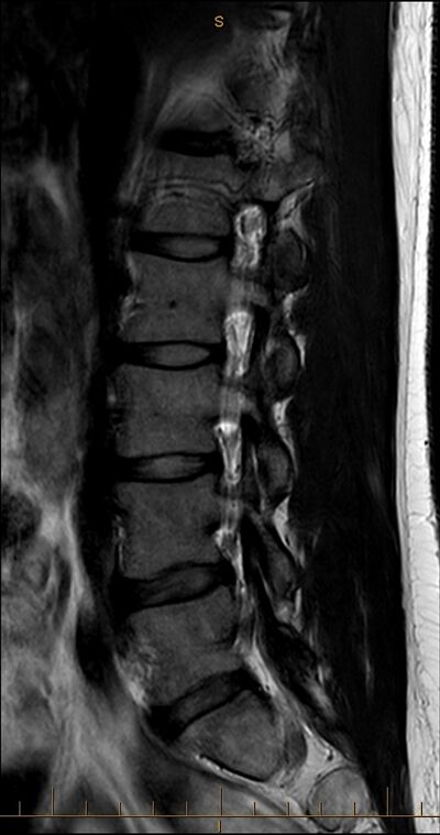
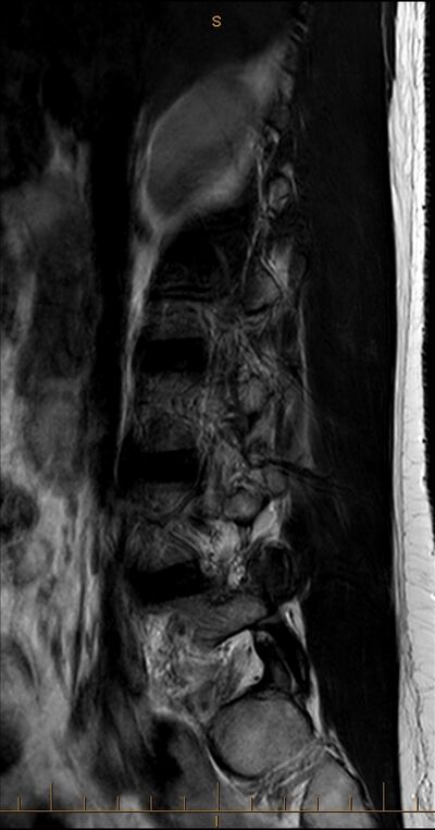
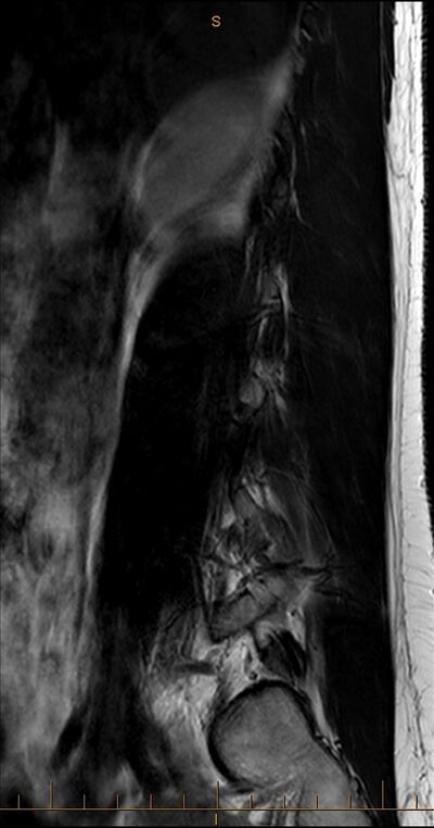
Sagittal MRI scans are typically taken across five standard planes. There may be further scans between these planes, or it may be slightly displaced to the left or right. | |||
{| class="wikitable" | |||
|+ Sagittal MRI five standard planes and the intersecting structures | |||
|- | |||
! Plane !! Vertebral structures !! Vertebral canal structures !! Extra-vertebral structures | |||
|- | |||
| style="width: 10%" |Median || style="width: 30%" | Vertebral bodies and intervertebral discs (AP and AF) anteriorly, and the spinous processes posteriorly || style="width: 30%" | Conus medullaris at upper segmental levels, and the cauda equina at lower levels. || Aorta terminates opposite L4 | |||
|- | |||
| Paramedian || Vertebral bodies and intervertebral discs anteriorly (AP and AF), and the laminae posteriorly || May miss the spinal cord, and only show the cauda equina. || Right of midline: right side edge of the aorta, right common iliac artery in front of L5, and left common iliac vein.<br>Left of midline passes through the aorta.<br>Posteriorly there is multifidus. | |||
|- | |||
| Transpedicular || Lateral sectors of the vertebral bodies, the lateral sectors of the intervertebral discs (AF +/- NP), the pedicles, and some portions of the facet joints. Slightly more medial intersects the inferiorly articular processes. Slightly more lateral intersects the superior articular processes || Spinal nerves and their dural sleeves as they pass under the pedicles in the intervertebral foraminae|| Right of midline: IVC, right common iliac artery in front of L5<br>Left of midline: might intersect lateral aorta<br>Posteriorly multifidus. | |||
|- | |||
| Tangential || Most lateral margins of the vertebral bodies and the AF at each segmental level, or the concavity of the vertebral body and their lumbar arteries and veins || No intersection || Left of midline: lateral to aorta or lateral segments<br>Right of midline: IVC, right common iliac artery in front of L5<br>Posteriorly the erector spinae at most levels and multifidus at lumbosacral levels. | |||
|- | |||
| Peripheral || Transverse processes || No intersection || Psoas major, quadratus lumborum anteriorly. Erector spinae posteriorly. | |||
|} | |||
*Anteriorly different planes have various intersections with the crura of the diaphragm, the great vessels, psoas major, and quadratus lumborum. | |||
*Posteriorly there is intersection through multifidus, longissimus thoracic, or iliocostalis lumborum. | |||
*Across the vertebral body concavities there are the lumbar arteries and lumbar veins that are covered by psoas | |||
*Anterior to the vertebral bodies on the right there is the right crus and the IVC | |||
*Anterior to the vertebral bodies towards the centre and left there is the left crus and the aorta | |||
*Posterior to the laminae there is multifidus | |||
*Anterior to the transverse processes there is the psoas major and quadratus lumborum | |||
*Posterior the the transverse processes there is various components of the erector spinae. | |||
{{Imagestack | |||
|width=400 | |||
|title=T2 Sagittal scans to the right of midline | |||
|align=centre | |||
|loop=no | |||
|File:MRI T2 Lumbar Spine Sagittal Median.jpg|Sagittal Median | |||
|File:MRI T2 Lumbar Spine Sagittal Paramedian.jpg|Sagittal Paramedian | |||
|File:MRI T2 Lumbar Spine Sagittal Transpedicular.jpg|Sagittal Transpedicular | |||
|File:MRI T2 Lumbar Spine Sagittal Tangential.jpg|Sagittal Tangential | |||
|File:MRI T2 Lumbar Spine Sagittal Peripheral.jpg|Sagittal Peripheral | |||
}} | |||
===Axial scans=== | |||
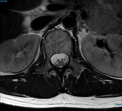
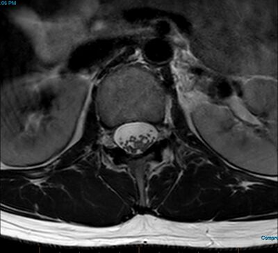
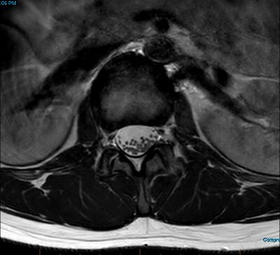
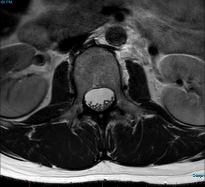
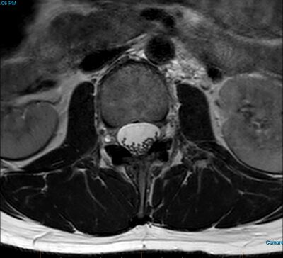
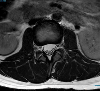
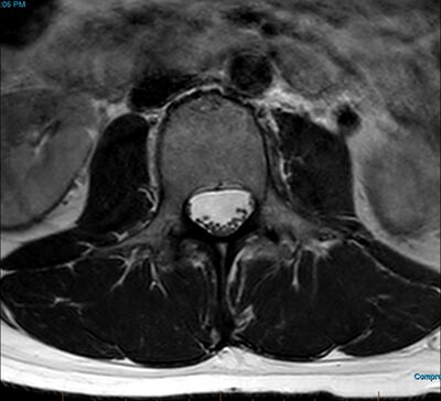
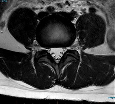
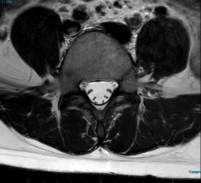
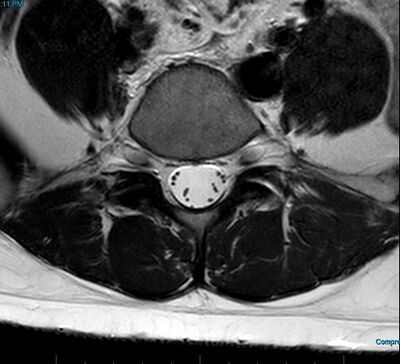
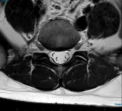
There are three main planes for axial MRI scans. With interpretation, the first step is determining whether the scan is transpedicular, subpedicular, or transarticular. The second step is estimating the segmental level by examining the external relations. | |||
{| class="wikitable" | |||
|+ Axial MRI three standard planes and the internal relations | |||
|- | |||
! Plane !! Vertebral structures !! Vertebral canal structures | |||
|- | |||
| style="width: 10%" |Transpedicular || style="width: 30%" | Pedicles project form the posterior surface of the vertebral body. Posteriorly various parts of the laminae and spinous process. || style="width: 30%" | Dural sac and nerve roots of the cauda equina. Nerve roots typically towards posterior surface of the sac due to the patient lying supine. | |||
|- | |||
| Subpedicular || Vertebral body anteriorly, laminae posteriorly || Dural sac with nerve roots of the cauda equina, and spinal nerves of the segment as they run through the intervertebral foramen. Spinal nerve lateral to the dural sac +/- dural sleeve with CSF. | |||
|- | |||
| Transarticular || Intervertebral disc, facet joints|| Dural sac and cauda equina centrally. Spinal nerve or ventral ramus leaving the vertebral column at the posterolateral corner of the disc and entering the psoas major. The spinal nerve passes obliquely and caudally through the foramen and so it is located further laterally than in subpedicular scans and is dissociated from the dural sac. | |||
|} | |||
The external relations can be used to identify the segment being used. | |||
{| class="wikitable" | |||
|+ External Relations | |||
|- | |||
! Segment !! Anterior || Lateral || Posterior | |||
|- | |||
| style="width: 10%" |L1 || style="width: 30%" |Left and right crus, IVC, Aorta, left renal vessels || style="width: 30%" |Broad flat quadratus lumborum, and a narrow psoas major || style="width: 30%" |Narrow multifidus plus longissimus thoracic, and iliocostalis lumborum. | |||
|- | |||
| L2 || Crura are smaller, IVC, Aorta, right renal vessels || Quadratus lumborum, larger psoas major || Multifidus, longissimus thoracic, and iliocostalis lumborum | |||
|- | |||
| L3 || No crura. IVC and Aorta || Quadratus lumborum, large psoas major || Multifidus expands laterally to the limits of the laminae. Plus longissimus thoracic and iliocostalis lumborum | |||
|- | |||
| L4 || 3 great vessels: IVC and common iliac arteries || large psoas major, small quadratus lumborum || Multifidus is wide, and erector spinae are small. | |||
|- | |||
| L5 || 4 great vessels: common iliac veins and arteries || Huge psoas major, absent quadratus lumborum, iliolumbar ligament || Multifidus is wide. No iliocostalis, and a small portion of longissimus. | |||
|} | |||
{{Imagestack | |||
|width=400 | |||
|title=T2 Axial scans | |||
|align=centre | |||
|loop=no | |||
|File:MRI T2 Lumbar Spine L1 Transpedicular Axial.jpg|L1 Transpedicular | |||
|File:MRI T2 Lumbar Spine L1 Subpedicular Axial.jpg|L1 Subpedicular | |||
|File:MRI T2 Lumbar Spine L1-L2 Transarticular Axial.jpg|L1-L2 Transarticular | |||
|File:MRI T2 Lumbar Spine L2 Transpedicular Axial.jpg|L2 Transpedicular | |||
|File:MRI T2 Lumbar Spine L2 Subpedicular Axial.jpg|L2 Subpedicular | |||
|File:MRI T2 Lumbar Spine L2-L3 Transarticular Axial.jpg|L2-L3 Transarticular | |||
|File:MRI T2 Lumbar Spine L3 Transpedicular Axial.jpg|L3 Transpedicular | |||
|File:MRI T2 Lumbar Spine L3 Subpedicular Axial.jpg|L3 Subpedicular | |||
|File:MRI T2 Lumbar Spine L3-L4 Transarticular Axial.jpg|L3-L4 Transarticular | |||
|File:MRI T2 Lumbar Spine L4 Transpedicular Axial.jpg|L4 Transpedicular | |||
|File:MRI T2 Lumbar Spine L4 Subpedicular Axial.jpg|L4 Subpedicular | |||
|File:MRI T2 Lumbar Spine L4-L5 Transarticular Axial.jpg|L4-L5 Transarticular | |||
|File:MRI T2 Lumbar Spine L5 Transpedicular Axial.jpg|L5 Transpedicular | |||
|File:MRI T2 Lumbar Spine L5 Subpedicular Axial.jpg|L5 Subpedicular | |||
|File:MRI T2 Lumbar Spine L5-S1 Transarticular Axial.jpg|L5-S1 Transarticular | |||
}} | |||
==Check List for Interpretation== | |||
Before doing anything, first verify patient identification and the date of scan | Before doing anything, first verify patient identification and the date of scan | ||
{{MRI Pulse Sequences}} | |||
===Survey View=== | ===Survey View=== | ||
| Line 16: | Line 113: | ||
*Neural foramina and nerve roots: nerve contact and compression. | *Neural foramina and nerve roots: nerve contact and compression. | ||
*Intervertebral discs: width, protrusions/ herniations. | *Intervertebral discs: width, protrusions/ herniations. | ||
*Spinal column: alignment (spondylolisthesis), vertebral body shape (compression fractures, Schmorls’ nodes), posterior bony elements (spondylolysis), degenerative end plate changes (changes in fat content), hemangiomas. | *Spinal column: alignment ([[spondylolisthesis]]), vertebral body shape (compression fractures, Schmorls’ nodes), posterior bony elements (spondylolysis), degenerative end plate changes (changes in fat content), hemangiomas. | ||
*Retroperitoneal space: adenopathy, masses, great vessel aneurysm, etc | *Retroperitoneal space: adenopathy, masses, great vessel aneurysm, etc | ||
| Line 25: | Line 122: | ||
*Dural sac—cord and rootlets: width, compression, irregularities | *Dural sac—cord and rootlets: width, compression, irregularities | ||
*Intervertebral discs: width, protrusions/ herniations, hydration, high intensity zones | *Intervertebral discs: width, protrusions/ herniations, hydration, high intensity zones | ||
*Spinal column: alignment (spondylolisthesis), vertebral body shape (compression fractures, Schmorls’ nodes), posterior bony elements (spondylolysis), degenerative end plate changes (changes in fat content), hemangiomas. | *Spinal column: alignment ([[spondylolisthesis]]), vertebral body shape (compression fractures, Schmorls’ nodes), posterior bony elements ([[spondylolysis]]), degenerative end plate changes (changes in fat content), hemangiomas. | ||
*Posterior bony elements: breakage, listhesis, pseudo-articulations, etc. | *Posterior bony elements: breakage, listhesis, pseudo-articulations, etc. | ||
| Line 34: | Line 131: | ||
*Content of the spinal canal and neural foramina: Trace course of nerve roots through neural foramina | *Content of the spinal canal and neural foramina: Trace course of nerve roots through neural foramina | ||
*Intervertebral discs— continuity, bulges, etc. | *Intervertebral discs— continuity, bulges, etc. | ||
*Bone – Vertebral bodies; spondylolisthesis, posterior bony elements (spondylolysis, breakage) | *Bone – Vertebral bodies; [[spondylolisthesis]], posterior bony elements ([[spondylolysis]], breakage) | ||
*Ligamentam flavum: thickened appearance, impingement | *Ligamentam flavum: thickened appearance, impingement | ||
*Retroperitoneal space: adenopathy, masses, muscle, fat infiltration of multifidus (see [[ | *Retroperitoneal space: adenopathy, masses, muscle, fat infiltration of multifidus (see [[:File:Multifidus fat infiltration.png|image]])<ref>{{#pmid:17254322}}</ref> | ||
===T2 Axials=== | ===T2 Axials=== | ||
| Line 42: | Line 139: | ||
*Content of the spinal canal and neural foramina: Trace course of nerve roots through neural foramina | *Content of the spinal canal and neural foramina: Trace course of nerve roots through neural foramina | ||
*Intervertebral discs— continuity, bulges, etc. | *Intervertebral discs— continuity, bulges, etc. | ||
*Bone – Vertebral bodies; spondylolisthesis, posterior bony elements (spondylolysis, breakage) | *Bone – Vertebral bodies; [[spondylolisthesis]], posterior bony elements ([[spondylolysis]], breakage) | ||
*Ligamentam flavum: thickened appearance, impingement | *Ligamentam flavum: thickened appearance, impingement | ||
*Retroperitoneal space: adenopathy, masses, muscle, etc. | *Retroperitoneal space: adenopathy, masses, muscle, etc. | ||
| Line 48: | Line 145: | ||
==Assessment== | ==Assessment== | ||
Ensure you have covered all structures. Assess need for other studies | Ensure you have covered all structures. Assess need for other studies | ||
==Bibliography== | |||
*Bogduk, Nikolai. Clinical and radiological anatomy of the lumbar spine. Chapter 19. Edinburgh: Elsevier/Churchill Livingstone, 2012. | |||
==References== | ==References== | ||
| Line 55: | Line 155: | ||
[[Category:Lumbar Spine]] | [[Category:Lumbar Spine]] | ||
[[Category:Radiology]] | [[Category:Radiology]] | ||
Latest revision as of 18:36, 20 October 2022
Anatomical Structures
Sagittal Scans
Sagittal MRI scans are typically taken across five standard planes. There may be further scans between these planes, or it may be slightly displaced to the left or right.
| Plane | Vertebral structures | Vertebral canal structures | Extra-vertebral structures |
|---|---|---|---|
| Median | Vertebral bodies and intervertebral discs (AP and AF) anteriorly, and the spinous processes posteriorly | Conus medullaris at upper segmental levels, and the cauda equina at lower levels. | Aorta terminates opposite L4 |
| Paramedian | Vertebral bodies and intervertebral discs anteriorly (AP and AF), and the laminae posteriorly | May miss the spinal cord, and only show the cauda equina. | Right of midline: right side edge of the aorta, right common iliac artery in front of L5, and left common iliac vein. Left of midline passes through the aorta. Posteriorly there is multifidus. |
| Transpedicular | Lateral sectors of the vertebral bodies, the lateral sectors of the intervertebral discs (AF +/- NP), the pedicles, and some portions of the facet joints. Slightly more medial intersects the inferiorly articular processes. Slightly more lateral intersects the superior articular processes | Spinal nerves and their dural sleeves as they pass under the pedicles in the intervertebral foraminae | Right of midline: IVC, right common iliac artery in front of L5 Left of midline: might intersect lateral aorta Posteriorly multifidus. |
| Tangential | Most lateral margins of the vertebral bodies and the AF at each segmental level, or the concavity of the vertebral body and their lumbar arteries and veins | No intersection | Left of midline: lateral to aorta or lateral segments Right of midline: IVC, right common iliac artery in front of L5 Posteriorly the erector spinae at most levels and multifidus at lumbosacral levels. |
| Peripheral | Transverse processes | No intersection | Psoas major, quadratus lumborum anteriorly. Erector spinae posteriorly. |
- Anteriorly different planes have various intersections with the crura of the diaphragm, the great vessels, psoas major, and quadratus lumborum.
- Posteriorly there is intersection through multifidus, longissimus thoracic, or iliocostalis lumborum.
- Across the vertebral body concavities there are the lumbar arteries and lumbar veins that are covered by psoas
- Anterior to the vertebral bodies on the right there is the right crus and the IVC
- Anterior to the vertebral bodies towards the centre and left there is the left crus and the aorta
- Posterior to the laminae there is multifidus
- Anterior to the transverse processes there is the psoas major and quadratus lumborum
- Posterior the the transverse processes there is various components of the erector spinae.
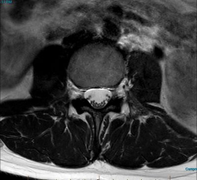
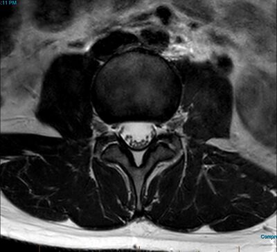
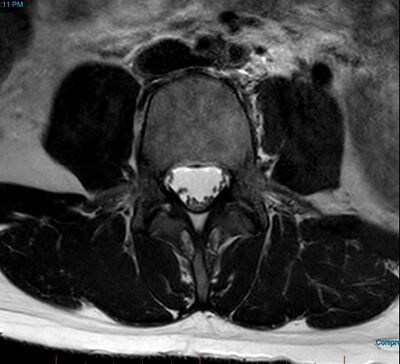
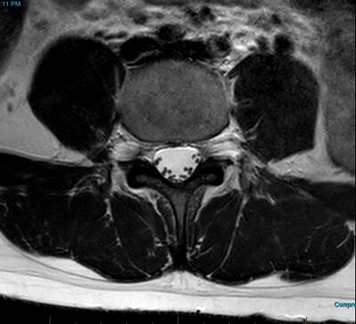
Axial scans
There are three main planes for axial MRI scans. With interpretation, the first step is determining whether the scan is transpedicular, subpedicular, or transarticular. The second step is estimating the segmental level by examining the external relations.
| Plane | Vertebral structures | Vertebral canal structures |
|---|---|---|
| Transpedicular | Pedicles project form the posterior surface of the vertebral body. Posteriorly various parts of the laminae and spinous process. | Dural sac and nerve roots of the cauda equina. Nerve roots typically towards posterior surface of the sac due to the patient lying supine. |
| Subpedicular | Vertebral body anteriorly, laminae posteriorly | Dural sac with nerve roots of the cauda equina, and spinal nerves of the segment as they run through the intervertebral foramen. Spinal nerve lateral to the dural sac +/- dural sleeve with CSF. |
| Transarticular | Intervertebral disc, facet joints | Dural sac and cauda equina centrally. Spinal nerve or ventral ramus leaving the vertebral column at the posterolateral corner of the disc and entering the psoas major. The spinal nerve passes obliquely and caudally through the foramen and so it is located further laterally than in subpedicular scans and is dissociated from the dural sac. |
The external relations can be used to identify the segment being used.
| Segment | Anterior | Lateral | Posterior |
|---|---|---|---|
| L1 | Left and right crus, IVC, Aorta, left renal vessels | Broad flat quadratus lumborum, and a narrow psoas major | Narrow multifidus plus longissimus thoracic, and iliocostalis lumborum. |
| L2 | Crura are smaller, IVC, Aorta, right renal vessels | Quadratus lumborum, larger psoas major | Multifidus, longissimus thoracic, and iliocostalis lumborum |
| L3 | No crura. IVC and Aorta | Quadratus lumborum, large psoas major | Multifidus expands laterally to the limits of the laminae. Plus longissimus thoracic and iliocostalis lumborum |
| L4 | 3 great vessels: IVC and common iliac arteries | large psoas major, small quadratus lumborum | Multifidus is wide, and erector spinae are small. |
| L5 | 4 great vessels: common iliac veins and arteries | Huge psoas major, absent quadratus lumborum, iliolumbar ligament | Multifidus is wide. No iliocostalis, and a small portion of longissimus. |
Check List for Interpretation
Before doing anything, first verify patient identification and the date of scan
- Basic Pulse Sequences for MRI[1]
| Image Type | TR | TE | Fat Signal Intensity | Water Signal Intensity | Advantages | Disadvantages |
|---|---|---|---|---|---|---|
| T1 | Short | Short | Bright | Dark | Best anatomic detail, rapid acquisition | Poor visualisation of pathology/oedema |
| T2 | Long | Long | Intermediate | Bright | Moderate sensitivity for pathology/oedema | Poor spatial resolution, time consuming |
| Fat-suppressed T2 | Long | Short | Very dark | Very bright | Most sensitive for pathology/oedema | Susceptible to artifacts related to magnetic resonance inhomogeneity |
| Gradient Echo | Short | Short | Intermediate | Intermediate/high | Excellent for articular cartilage, PVNS, and blood | Very susceptible to metallic artifacts |
| Proton Density | Long | Short | Intermediate/high | Intermediate | Excellent for meniscal pathology |
Survey View
Count vertebra, and assess for transitional anatomy
Fat Suppressed Coronal
Assess facet joints for effusions, size, cartilage thickness, oedema. Look for extra spinal pathology. Scroll through the sacroiliac joints looking for osteoarthritis, oedema, effusions.
T1 Sagittals
In T1, spinal fluid is dark and fat is bright. Determine the left-right orientation. On the left, the gives off branches at ~L1. On the right, the renal artery runs posterior to IVC. The Aorta is wider than IVC.
Working from caudal to rostral observe:
- Neural foramina and nerve roots: nerve contact and compression.
- Intervertebral discs: width, protrusions/ herniations.
- Spinal column: alignment (spondylolisthesis), vertebral body shape (compression fractures, Schmorls’ nodes), posterior bony elements (spondylolysis), degenerative end plate changes (changes in fat content), hemangiomas.
- Retroperitoneal space: adenopathy, masses, great vessel aneurysm, etc
T2 Sagittals
In T2, spinal fluid is bright.
Working from caudal to rostral observe:
- Dural sac—cord and rootlets: width, compression, irregularities
- Intervertebral discs: width, protrusions/ herniations, hydration, high intensity zones
- Spinal column: alignment (spondylolisthesis), vertebral body shape (compression fractures, Schmorls’ nodes), posterior bony elements (spondylolysis), degenerative end plate changes (changes in fat content), hemangiomas.
- Posterior bony elements: breakage, listhesis, pseudo-articulations, etc.
T1 Axials
The CSF appears gray and fat appears bright. Proceed caudal to cranial.
Orientation – neural foramina lie at level of discs.
- Content of the spinal canal and neural foramina: Trace course of nerve roots through neural foramina
- Intervertebral discs— continuity, bulges, etc.
- Bone – Vertebral bodies; spondylolisthesis, posterior bony elements (spondylolysis, breakage)
- Ligamentam flavum: thickened appearance, impingement
- Retroperitoneal space: adenopathy, masses, muscle, fat infiltration of multifidus (see image)[2]
T2 Axials
Spinal fluid appears bright. Proceed caudal to cranial.
- Content of the spinal canal and neural foramina: Trace course of nerve roots through neural foramina
- Intervertebral discs— continuity, bulges, etc.
- Bone – Vertebral bodies; spondylolisthesis, posterior bony elements (spondylolysis, breakage)
- Ligamentam flavum: thickened appearance, impingement
- Retroperitoneal space: adenopathy, masses, muscle, etc.
Assessment
Ensure you have covered all structures. Assess need for other studies
Bibliography
- Bogduk, Nikolai. Clinical and radiological anatomy of the lumbar spine. Chapter 19. Edinburgh: Elsevier/Churchill Livingstone, 2012.
References
- ↑ Khanna et al.. Magnetic resonance imaging of the knee. Current techniques and spectrum of disease. The Journal of bone and joint surgery. American volume 2001. 83-A Suppl 2 Pt 2:128-41. PMID: 11712834. DOI.
- ↑ Kjaer et al.. Are MRI-defined fat infiltrations in the multifidus muscles associated with low back pain?. BMC medicine 2007. 5:2. PMID: 17254322. DOI. Full Text.
Literature Review
- Reviews from the last 7 years: review articles, free review articles, systematic reviews, meta-analyses, NCBI Bookshelf
- Articles from all years: PubMed search, Google Scholar search.
- TRIP Database: clinical publications about evidence-based medicine.
- Other Wikis: Radiopaedia, Wikipedia Search, Wikipedia I Feel Lucky, Orthobullets,
