Lumbar Spine MRI
Interpretation
Before doing anything, first verify patient identification and the date of scan
Survey View
Count vertebra, and assess for transitional anatomy
Fat Suppressed Coronal
Assess facet joints for effusions, size, cartilage thickness, oedema. Look for extra spinal pathology. Scroll through the sacroiliac joints looking for osteoarthritis, oedema, effusions.
T1 Sagittals
In T1, spinal fluid is dark and fat is bright. Determine the left-right orientation. On the left, the gives off branches at ~L1. On the right, the renal artery runs posterior to IVC. The Aorta is wider than IVC.
Working from caudal to rostral observe:
- Neural foramina and nerve roots: nerve contact and compression.
- Intervertebral discs: width, protrusions/ herniations.
- Spinal column: alignment (spondylolisthesis), vertebral body shape (compression fractures, Schmorls’ nodes), posterior bony elements (spondylolysis), degenerative end plate changes (changes in fat content), hemangiomas.
- Retroperitoneal space: adenopathy, masses, great vessel aneurysm, etc
T2 Sagittals
In T2, spinal fluid is bright.
Working from caudal to rostral observe:
- Dural sac—cord and rootlets: width, compression, irregularities
- Intervertebral discs: width, protrusions/ herniations, hydration, high intensity zones
- Spinal column: alignment (spondylolisthesis), vertebral body shape (compression fractures, Schmorls’ nodes), posterior bony elements (spondylolysis), degenerative end plate changes (changes in fat content), hemangiomas.
- Posterior bony elements: breakage, listhesis, pseudo-articulations, etc.
T1 Axials
The CSF appears gray and fat appears bright. Proceed caudal to cranial.
Orientation – neural foramina lie at level of discs.
- Content of the spinal canal and neural foramina: Trace course of nerve roots through neural foramina
- Intervertebral discs— continuity, bulges, etc.
- Bone – Vertebral bodies; spondylolisthesis, posterior bony elements (spondylolysis, breakage)
- Ligamentam flavum: thickened appearance, impingement
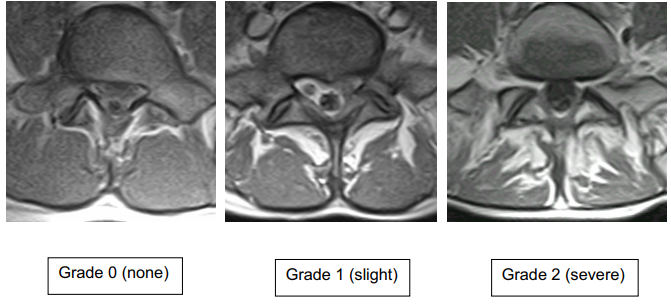
- Retroperitoneal space: adenopathy, masses, muscle, fat infiltration of multifidus (see image)[1]
T2 Axials
Spinal fluid appears bright. Proceed caudal to cranial.
- Content of the spinal canal and neural foramina: Trace course of nerve roots through neural foramina
- Intervertebral discs— continuity, bulges, etc.
- Bone – Vertebral bodies; spondylolisthesis, posterior bony elements (spondylolysis, breakage)
- Ligamentam flavum: thickened appearance, impingement
- Retroperitoneal space: adenopathy, masses, muscle, etc.
Assessment
Ensure you have covered all structures. Assess need for other studies
References
Literature Review
 Literature Review
- Reviews from the last 7 years: review articles, free review articles, systematic reviews, meta-analyses, NCBI Bookshelf
- Articles from all years: PubMed search, Google Scholar search.
- TRIP Database: clinical publications about evidence-based medicine.
- Other Wikis: Radiopaedia, Wikipedia Search, Wikipedia I Feel Lucky, Orthobullets,

