Metacarpal Fractures: Difference between revisions
mNo edit summary |
No edit summary |
||
| (4 intermediate revisions by the same user not shown) | |||
| Line 1: | Line 1: | ||
{{ | {{Creative commons not compatible|source=[https://orthopaedia.com/page/Metacarpal-fractures Orthopaedia.com]|terms=not for commercial use, but can be modified.|license=[https://creativecommons.org/licenses/by-nc-sa/4.0/ CC-BY-NC-SA license]}} | ||
The metacarpals are essential for hand function. Fractures to these bones may affect hand strength and motion, inhibiting the ability to grip and hold objects. Fractures are often the result of high-energy impact, likely seen in athletics, trauma and work injuries. These fractures often can be treated with immobilization and have a good prognosis. | The metacarpals are essential for hand function. Fractures to these bones may affect hand strength and motion, inhibiting the ability to grip and hold objects. Fractures are often the result of high-energy impact, likely seen in athletics, trauma and work injuries. These fractures often can be treated with immobilization and have a good prognosis. | ||
== Structure and Function == | == Structure and Function == | ||
[[File:Metacarpal bones.jpg|thumb|'''Figure 1''': The 5 metacarpal bones are shown in red.]] | [[File:Metacarpal bones.jpg|thumb|'''Figure 1''': The 5 metacarpal bones are shown in red.]]{{See also|Hand and Wrist Biomechanics}} | ||
The metacarpals consist of five tubular bones that articulate proximally with the carpus and distally with the phalanges (Figure 1). From radial to ulnar, the bones are numbered one (thumb) through five (small finger) and consist of a shaft, base, and head. | The metacarpals consist of five tubular bones that articulate proximally with the carpus and distally with the phalanges (Figure 1). From radial to ulnar, the bones are numbered one (thumb) through five (small finger) and consist of a shaft, base, and head. | ||
| Line 17: | Line 17: | ||
== Patient Presentation == | == Patient Presentation == | ||
Metacarpal fractures usually occur after a fistfight, car accident, or fall. At times, a crush mechanism is responsible. Findings include pain (most intense over fracture site), oedema, a shortened finger or finger deformity (such as depressed or missing knuckle), and bruising. | Metacarpal fractures usually occur after a fistfight, car accident, or fall. At times, a crush mechanism is responsible. Findings include pain (most intense over fracture site), oedema, a shortened finger or finger deformity (such as depressed or missing knuckle), and bruising. | ||
| Line 39: | Line 34: | ||
Thumb metacarpal shaft fractures are treated similarly to the other four metacarpals. | Thumb metacarpal shaft fractures are treated similarly to the other four metacarpals. | ||
<gallery heights=300px widths=300px> | |||
Fracture base of 5th metacarpal.jpg|'''Figure 2''': Fracture of the base of the 5th metacarpal. Soft tissue swelling, denoted by the arrow, is perhaps a more obvious finding than the fracture itself. To the right, a close-up view of the base of the metacarpal shows the fracture line, outlined in red in the image below. | |||
Fracture shaft 5th metacarpal.jpg|'''Figure 3:''' Fracture of the shaft of the 5th metacarpal. | |||
Boxers fracture.jpg|'''Figure 4''': Boxer's fracture, i.e., a fracture of the neck of the 5th metacarpal. First metacarpal (thumb) fractures are uncommon. However, these fractures deserve special attention. | |||
Bennetts fracture.jpg|'''Figure 5''': Bennett's fracture. | |||
Rolandos fracture.jpg|'''Figure 6''': Rolando's fracture. | |||
</gallery> | |||
== Objective Evidence == | == Objective Evidence == | ||
Latest revision as of 06:02, 2 April 2022
The metacarpals are essential for hand function. Fractures to these bones may affect hand strength and motion, inhibiting the ability to grip and hold objects. Fractures are often the result of high-energy impact, likely seen in athletics, trauma and work injuries. These fractures often can be treated with immobilization and have a good prognosis.
Structure and Function

- See also: Hand and Wrist Biomechanics
The metacarpals consist of five tubular bones that articulate proximally with the carpus and distally with the phalanges (Figure 1). From radial to ulnar, the bones are numbered one (thumb) through five (small finger) and consist of a shaft, base, and head.
The metacarpal base is cuboidal in shape, and ligaments provide articulations between the metacarpal bones and the carpus at the carpometacarpal (“CMC”) joint.
The body (or shaft) of the metacarpal is concave with respect to the palmar surface. There is a medial, lateral, and dorsal surface on the metacarpal shaft. The medial and lateral surfaces provide attachment for the interossei muscles. This attachment, along with the extensor and flexor muscles crossing (but not attaching to the metacarpal), produces the main deforming force that may displace shaft fractures. The extensor tendons lie atop the flat dorsum of the metacarpal shaft.
The metacarpal neck lies just proximal to the head, distal to the shaft.
The metacarpal head articulates with the proximal phalanx of each finger, with tubercles on each side providing attachment for the collateral ligaments. The dorsum of the head accommodates the extensor tendons while the palmar surface has a ridge for the flexor tendons. The head of the metacarpal receives its own blood supply from the collateral ligaments; this arrangement predisposes the head to possible avascular necrosis with a ligament injury, as the ligament injury may disrupt perfusion.
The first metacarpal is shorter and wider than the other metacarpals and has a more extreme angulation with the carpus. The second and third metacarpals are more rigidly fixed upon the corresponding carpal bones than the fourth and fifth metacarpals. The looser attachments of the fourth and fifth metacarpals allow them to oppose the thumb.
Patient Presentation
Metacarpal fractures usually occur after a fistfight, car accident, or fall. At times, a crush mechanism is responsible. Findings include pain (most intense over fracture site), oedema, a shortened finger or finger deformity (such as depressed or missing knuckle), and bruising.
Physical examination of the hand may reveal deformities, decreased grip strength, and possible finger misalignment. In a normal flexed hand, the fingertips should align and point towards the scaphoid tubercle on the radial volar aspect of the wrist. Range of motion tests should be conducted, and the hand should be assessed for sensory changes to determine possible nerve damage.
Fractures within each of the four regions of the metacarpal bone – base (Figure 2), shaft (Figure 3), neck, and head – are considered distinctly.
Metacarpal base fractures usually occur as a result of significant axial load to a flexed hand as seen in clenched fist injuries.
Shaft fractures are most frequently the result of direct trauma, axial loading, or twisting injury. These present respectively as transverse fractures (with comminution at times) and spiral fractures. The pull of the interossei muscles and flexor tendons can deform shaft fractures leading to metacarpal shortening or angulation.
Metacarpal neck fractures are the most common type of metacarpal fracture. Such a fracture seen in the 5th (or rarely, the 4th) metacarpal neck is called a "boxer’s fracture" (Figure 4). Metacarpal head fractures are rare and usually require surgical intervention.
A notable thumb injury is the Bennett's fracture (Figure 5), a partial articular fracture of the first metacarpal base. This fracture results in shortening and deformation of the thumb, due to lack of support from the bone and shear pull from the abductor pollicis longus.
Another notable thumb injury is the Rolando's fracture (Figure 6), one with T or Y shaped intra-articular fracture line at the base of the thumb metacarpal. This fracture is similar to the Bennett's fracture, but with more comminution and shortening; it is, therefore, a more severe injury.
Thumb metacarpal shaft fractures are treated similarly to the other four metacarpals.
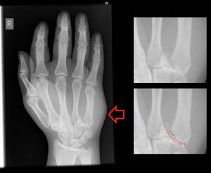
Figure 2: Fracture of the base of the 5th metacarpal. Soft tissue swelling, denoted by the arrow, is perhaps a more obvious finding than the fracture itself. To the right, a close-up view of the base of the metacarpal shows the fracture line, outlined in red in the image below.
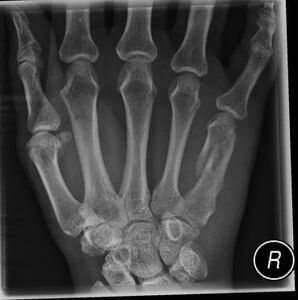
Figure 3: Fracture of the shaft of the 5th metacarpal.
Figure 4: Boxer's fracture, i.e., a fracture of the neck of the 5th metacarpal. First metacarpal (thumb) fractures are uncommon. However, these fractures deserve special attention.
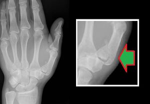
Figure 5: Bennett's fracture.
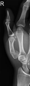
Figure 6: Rolando's fracture.


Objective Evidence
The following three X-ray views should be obtained in order to confirm a metacarpal fracture: posteroanterior (PA), oblique, and lateral. If a metacarpal head fracture is suspected but not seen well, special views (e.g., Brewerton view, in which the patient's hand is flexed at the metacarpophalangeal joint to 65 degrees) or CT scanning may be helpful.
Epidemiology
Metacarpal fractures represent 10% of all fractures, and there is a lifetime incidence rate of 2.5%. The fifth metacarpal is fractured most frequently and accounts for one-fourth of all metacarpal fractures. Most of these fractures occur in the young, active, or working population and consequently result in a significant loss of revenue and work time each year.
Differential Diagnosis
The differential diagnosis of metacarpal fractures can be thought of as “bad,” “not so bad,” and “very bad.” A non-displaced shaft fracture may be “not so bad;” an angulated fracture sustained by punching a fellow in the face, with a resultant open wound could easily be “very bad.” To sort that out, the following features of the injury must be delineated:
- The location of the injury (which bone, which region and articular involvement).
- The pattern of the fracture (e.g. simple vs comminuted; transverse vs spiral).
- Degree of displacement and rotational misalignment.
- Status of the soft tissue, paying special attention to possible fight bite wounds.
Red Flags
Malrotation of the fingers. Assessments of the patient’s hand rotation can be made by asking the patient to fold their hand half way into a fist. The nails should align in parallel, facing the ceiling; fingertips should point to the scaphoid. The key is that rotation cannot be determined by examining an x-ray. It must be noted during the physical examination.
A punch against the face is at risk for a fight bite – a small wound that is replete with mouth flora (and subsequent infection). The bite mark may not be located near the metacarpophalangeal joint, but the injury may still have damaged the joint capsule. Look closely for this, as it requires prompt surgical intervention.
“Trivial” chip fractures may represent avulsions of the ligaments and a significant soft tissue injury. The size of the bone fragment does not necessarily correlate with the severity of the injury.
Profound hand swelling from a crush injury may produce a compartment syndrome.
Treatment Options and Outcomes

The three aims of treatment are preserving alignment, mobility and proper finger length.
The following factors should be accounted for to determine whether surgical (open reduction) or non-surgical (closed reduction) intervention is best:
- degree of angulation
- amount of displacement
- presence of excessive rotation
Other factors that might be considered are the patient's age and occupational/recreational demands.
The guidelines regarding how much angulation is tolerable has more to do with eminence-based medicine than evidence-based medicine. The medical eminences have said that up 10 degrees of angulation can be accepted in the 2nd metacarpal with an additional 10 degrees for every finger moving in the ulnar direction, i.e., 3rd = 20 degrees; 4th = 30 degrees; and 5th = 40 degrees.
And speaking of “eminences”: after a fracture, the prominence of the 5th metacarpal-phalangeal joint is apt to be lost – a “cosmetic,” not functional loss — with closed treatment. Patients should be so warned.
If surgery is not needed, the patient's fracture should remain immobilized and they should be seen for follow-up in one to two weeks. X-rays should once again be taken to determine if there is proper healing and ensure no further displacement or angulation.
Immobilization of a metacarpal fracture must include at least the proximal phalanx, but there is no particular need to immobilize the inter-phalangeal joints and indeed that may lead to unnecessary stiffness. The metacarpal-phalangeal joint should be held in full flexion as long as the position doesn't further displace the fracture. Such a position will make the collateral ligaments maximally taut (because of the cam shape of the metacarpal head). This can help reduce the distal fracture fragment. This position can also prevent contractures of the joint. (If the ligament were not immobilized under maximal tension, it might set in this shorter position and lose its ability to stretch; subsequently, a joint contracture might form.) Within the cast, the affected finger should be buddy-taped to its neighbor. A vast majority of metacarpal neck fractures affect either the 4th or the 5th metacarpal. In those cases, the 2nd and 3rd fingers can be left free, to fully extend and flex.
When surgery is needed, options include pins, screws, metal wires, and plates (Figure 7). Open reduction of metacarpal head injuries demand an especially delicate touch, to avoid disruption of the blood supply and prevent avascular necrosis.
If only casting is needed, recovery should take about six to eight weeks. After three or four weeks of immobilization, the cast may gradually be removed and replaced with a removable splint. Radiographic reassessment should occur one week following the incident and at three week intervals after that. Limited range of motion exercises may be started after three weeks.
If surgical intervention is needed, recovery should take a few months, depending on the stability of the fracture and the care the patient takes to protect the hand from re-injury. The implants will remain in the hand unless they become bothersome to the patient.
In general, with proper treatment and rehabilitation, the prognosis for a metacarpal fracture is excellent. Bone healing begins around six weeks, but it may take years to return to full strength.
The duration of closed treatment has to be “just right.” Left on too long, a cast can cause joint contractures, yet casting for an inadequate period increases the risk for displacement and malunion.
Risk Factors and Prevention
Athletes who participate in sports, such as martial arts and football, have an increased risk of metacarpal fracture. Boxers frequently make closed fist contact with solid objects, while football players’ hands are frequently exposed and unprotected, which may lead to high energy impact and crush injuries.
Males are three times more likely than females to experience a metacarpal fracture.
Furthermore, adolescents, those in the work force, and the physically active populations have a greater risk of metacarpal injuries.
Miscellaneous
A shortened fourth metacarpal may signify congenital XO abnormality (Turner's syndrome).
Boxer's fractures in non-boxers may be a sign of anger management issues and possible domestic violence (especially when the recited history is “I fell”).
A boxer’s fracture may suggest not only trouble controlling one’s temper but poor boxing technique: ideally, the force of a punch should be transmitted via the more rigid 2nd and 3rd metacarpals. But don't tease the patient – he may slug you with the good hand.
Skills and Competencies
Be able to communicate orally the nature of the injury. Recognize “fight bite” fractures. Splint the fracture. Recognize which fractures might need surgical stabilization.
References
Part or all of this article or section is derived from Metacarpal Fractures by Orthopaedia.com, used under CC-BY-NC-SA

