Nerves of the Lumbar Spine
From WikiMSK
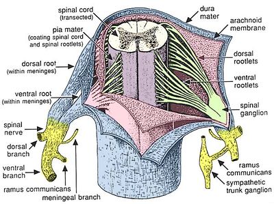
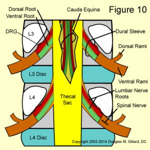
The lumbar spinal nerves lie in the intervertebral foraminae, and are connected by spinal nerve roots to the spinal cord. The spinal nerves divide into the ventral and dorsal rami outside the vertebral column. The lumbar sympathetic trunks run along the anterolateral lumbar vertebral column, and they communicate with the ventral rami of the lumbar spinal nerves
Lumbar Spinal Nerves
- The lumbar spinal nerves are numbered according to the vertebra beneath which they lie. The L1 spinal nerve lies below the L1 vertebra in the L1/2 intervertebral foramen. The L2 spinal nerve lies below the L2 vertebra in the L2/3 intervertebral foramen etc.
- Each spinal nerve is connected centrally to the spinal cord by a dorsal and ventral root.
- The joining of the spinal nerve roots to the spinal nerve occurs in the intervertebral foramen.
- The spinal nerve divides peripherally into a larger ventral ramus and smaller dorsal ramus.
- The branching into the two rami occurs outside the foramen.
- The spinal nerves are short, and no longer than the width of their intervertebral foramen.
- They are generally a few millimetres, but can be less than 1mm with the roots branching directly into rami without the formation of a proper spinal nerve.
Lumbar Nerve Roots
- The dorsal root transmits sensory fibres from the spinal nerve to the spinal cord
- The ventral root transmits mostly motor fibres from the spinal cord to the spinal nerve
- The ventral root may also transmit some sensory fibres
- The L1 and L2 ventral roots also transmit preganglionic, sympathetic, efferent fibres
- The spinal cord has its termination opposite the L1/2 intervertebral disc, but the range is T12/L1 to L2/3.
- The lower lumbar and sacral nerve roots run within the vertebral canal largely enclosed in the dural sac.
- In the cauda equina, the lumbar, sacral, and coccygeal nerve roots run freely
- Each root is covered by individual pia mater, continuous with the pia mater of the spinal cord.
- The roots of the cauda equina are within the cerebrospinal fluid (CSF) which flows through the subarachnoid space
- The nerve fibres within the nerve roots run in a single trunk for most of their course
- Rootlets
- Near the spinal cord they separate into rootlets which are smaller bundles, and attach to the spinal cord.
- There are between 2-12 rootlets for each root, and are 0.5-1mm in diameter.
- The ventral root rootlets attach to the ventrolateral aspect of the spinal cord
- The dorsal root rootlets attach to the dorsolateral sulcus of the cord and along the ventral and dorsal surface of the cord the rootlets form an uninterrupted series of attachments
- Dural sleeve
- A pair of spinal nerve roots leaves the dural sac just above the level of the intervertebral foramen
- The nerve roots penetrate the dural sac inferolaterally
- They take an extension of dura mater and arachnoid mater called the dural sleeve
- The dural sleeve encloses the nerve roots merges or becomes the epineurium of the spinal nerve
- The nerve roots are sheathed with pia mater and CSF flows around them as far as the spinal nerve
- Dorsal root ganglion
- The dorsal root ganglion is an enlargement of the dorsal root
- It is formed immediately proximal to the dorsal roots junction with the spinal nerve.
- It contains the cell bodies of the sensory fibres in the dorsal root
- It lies within the dural sleeve of the nerve roots and occupies the upper, medial part of the intervertebral foramen. It may lie further distally in the foramen with short spinal nerves.
- Nerve root angles leaving the dural sac
- The angles of each pair of nerve root as it leaves the dural sac varies, getting increasingly acute angles more caudally.
- The L1 nerve root leaves at about 80°, the L2 at about 70°, L3 and L4 at 60°, and L5 at 45°.
- Nerve root vertebral body origins
- The nerve root sleeves generally arise opposite the back of their respective vertebral bodies (L1 sleeve arises behind the L1 body, etc)
- However with successively caudal sleeves, they arise increasingly higher behind their vertebral bodies.
- And so the L5 nerve root arises behind the L4/5 intervertebral disc.
Relations of the Nerve Roots
Anomalies of the Nerve Roots
Dorsal Rami
Ventral Rami
Dermatomes
- Main article: Dermatomes
Sympathetic Nerves
Sinuvertebral Nerves
Innervation of the Lumbar Intervertebral Discs
Resources
See this brilliant visual tour of the lumbar nerve roots by a US physiotherapist.
References
These are study notes taken from Chapter 10 of:
- Bogduk, Nikolai. Clinical and radiological anatomy of the lumbar spine. Edinburgh: Elsevier/Churchill Livingstone, 2012.


