Proximal Weakness
Proximal muscle weakness involves the axial and limb girdle muscle groups and can be caused by a wide array of different neuromuscular diseases. Patients with this findings may have difficulty flexing or extending their neck against resistance, which can be detected by observing the patient sitting up from the supine position. In severe cases, patients may experience difficulty or be unable to sit up, and this may be the only objective evidence of their weakness.
Classification
| Proximal Weakness | |||||||||||||||||||||||||||||||||
| Myopathic | Non-Myopathic | ||||||||||||||||||||||||||||||||
| Acquired | Genetic | ||||||||||||||||||||||||||||||||
|
|
| |||||||||||||||||||||||||||||||
Examination

Assess for contractures, atrophy, and hypertrophy; seen in various disorders that cause proximal weakness
Lower limbs
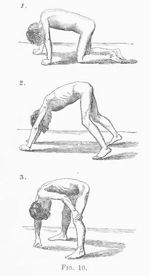
Gower's sign is a physical finding used to assess for proximal pelvic girdle muscle weakness particularly in those with certain neuromuscular disorders.[1] Gower thought it was pathognomonic for what is now called Duchenne muscular dystrophy, but it can acutally be seen in a variety of condition causing proximal weakness.
To perform the Gower's sign test, the patient is asked to rise from a sitting or lying position on the floor without using their hands or arms for support. Patients with normal muscle strength are able to perform this task with ease. However, patients with proximal lower limb muscle may use their hands and arms to "climb up" their own body, pushing against their thighs, hips, and abdomen to achieve a standing position. When rising from lying patients first turn prone before standing up. This maneuver is often accompanied by compensatory movements, such as hyperextension of the knees or lordosis of the lumbar spine.
This position helps reduce the amount of force required for knee extension and also facilitates the co-activation of multiple muscle groups during the standing-up process. When extending the knee, the hamstring muscles also contract to achieve maximum extension. Additionally, upper extremity abduction and extension helps with hip extension. When the gluteus medius and minimus muscles are weak, a wide hip abduction compensates by providing a more stable base. In cases of severe weakness, hand support may be necessary, and heel contracture and equine posturing may be present. However, when the equine becomes too severe or when contractures develop, external support may be needed to stand up. In such cases, patients use their hands to push on their knees and lift their trunks in a process referred to as "climbing up oneself."
Patients may also experience sudden dropping into a chair when attempting to sit down slowly.
Upper Limbs
For the upper limbs there may be scapular winging and difficulty in abduction.
Deltoid muscle strength can be evaluated by pressing down on fully abducted arms with the elbows flexed, and normal strength is indicated when the examiner is unable to overcome the patient's resistance.
Investigations

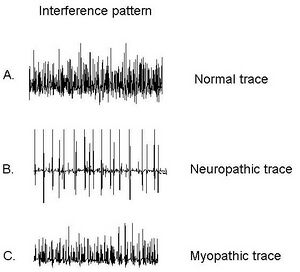
Electromyography can be particularly helpful in differentiating between myopathic and neuropathic causes. In a normal EMG trace, the electrical activity of a muscle appears as a smooth, regular pattern of muscle activation. In a neuropathic EMG trace, the electrical activity of the muscle appears abnormal, with irregular patterns of muscle activation. In a myopathic EMG trace, the electrical activity of the muscle appears increased and often shows signs of rapid firing or muscle fibre recruitment.
Resources
EURO-NMD Lecture on Proximal Weakness
References
- ↑ 1.0 1.1 Gowers. Pseudo-hypertrophic muscular paralysis : a clinical lecture. 1879. From https://archive.org/details/b21517034/page/n13/mode/2up
- ↑ Wu, Yunfen; Martnez Martnez, Mara ngeles; Orizaola Balaguer, Pedro (2013-05-22). Turker, Dr.Hande (ed.). "Overview of the Application of EMG Recording in the Diagnosis and Approach of Neurological Disorders" (in English). InTech. doi:10.5772/56030. ISBN 978-953-51-1118-4. Cite journal requires
|journal=(help)
Literature Review
- Reviews from the last 7 years: review articles, free review articles, systematic reviews, meta-analyses, NCBI Bookshelf
- Articles from all years: PubMed search, Google Scholar search.
- TRIP Database: clinical publications about evidence-based medicine.
- Other Wikis: Radiopaedia, Wikipedia Search, Wikipedia I Feel Lucky, Orthobullets,


