Quadratus Femoris Injection
| Quadratus Femoris Injection | |
|---|---|
| Indication | Ischiofemoral Impingement Syndrome |
Anatomy

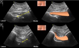
The ischiofemoral space is bounded by the ischial tuberosity medially, the lesser trochanter of the femur laterally, the femoral neck, ischiofemoral ligament, and inferior gemellus muscle cranially, and the inferior edge of the quadratus femoris muscle caudally. This quadrangular muscle originates from the ischial tuberosity and attaches to the femur's intertrochanteric crest. It is innervated by the nerve to the quadratus femoris (L4-S1) and supplied by the inferior gluteal artery.
Beneath the quadratus femoris lies the obturator externus muscle, which originates from the obturator membrane and bony boundaries of the obturator foramen and inserts into the femur's trochanteric fossa. It is innervated by the obturator nerve (L3-L4) and supplied by the obturator and medial femoral circumflex arteries. Both muscles are commonly implicated in ischiofemoral impingement syndrome (IIS).
Notably, the semitendinosus, semimembranosus, and biceps femoris muscles' conjoint tendon has proximal attachments on the ischial tuberosity, and their pathologies can be difficult to distinguish from IIS. Additionally, the sciatic and inferior gluteal nerves traverse the ischiofemoral space, and their entrapment may cause sciatica-like symptoms.
Indications and Efficacy
Suspicion for Ischiofemoral Impingement Syndrome. The procedure can be diagnostic and/or therapeutic in nature. There are limited data on efficacy.
Technique

Ultrasound Guided
The following technique has been described:[2]
- Patient Position: Prone
- Ultrasound Technique: use an 8-4 MHz curvilinear probe in a transverse or axial plane
- Start with ischial tuberosity as initial bony landmark
- Identify hamstring origin in soft-tissue
- Move transducer laterally to find lesser trochanter
- Note gluteus maximus muscle overlaying bony structures
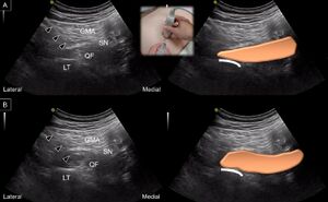
- Visualize quadratus femoris muscle which lies below the sciatic nerve
- Sciatic nerve usually near and lateral to hamstring tendon origin
- Needle Approach: lateral to medial, in-plane with transducer, both laterally and deep relative to sciatic nerve
- Visualize needle tip and sciatic nerve in real time
- Place needle tip in quadratus femoris muscle
- Needle Type: 3.5-inch (8.9 cm), 22-gauge spinal needle
- Injection Solution: 4 mL, containing 1 mL of triamcinolone and 3 mL of 1% lidocaine
An obliquely oriented path with the target in the lower third of the muscle may be the safest and less painful.
Complications
Puncture of quadratus, sciatic and posterior femoral cutaneous nerves, and posterior circumflex artery.
Aftercare
Assess diagnostic response
Videos
https://www.youtube.com/watch?v=-UvZOsrETfY
See Also
External Links
References
- ↑ 1.0 1.1 Wu, Wei-Ting; Chang, Ke-Vin; Mezian, Kamal; Naňka, Ondřej; Ricci, Vincenzo; Chang, Hsiang-Chi; Wang, Bow; Hung, Chen-Yu; Özçakar, Levent (2022-12-31). "Ischiofemoral Impingement Syndrome: Clinical and Imaging/Guidance Issues with Special Focus on Ultrasonography". Diagnostics (Basel, Switzerland). 13 (1): 139. doi:10.3390/diagnostics13010139. ISSN 2075-4418. PMC 9818255. PMID 36611431.
- ↑ Backer, Matthew W.; Lee, Kenneth S.; Blankenbaker, Donna G.; Kijowski, Richard; Keene, James S. (2014-09). "Correlation of ultrasound-guided corticosteroid injection of the quadratus femoris with MRI findings of ischiofemoral impingement". AJR. American journal of roentgenology. 203 (3): 589–593. doi:10.2214/AJR.13.12304. ISSN 1546-3141. PMID 25148161. Check date values in:
|date=(help)
Literature Review
- Reviews from the last 7 years: review articles, free review articles, systematic reviews, meta-analyses, NCBI Bookshelf
- Articles from all years: PubMed search, Google Scholar search.
- TRIP Database: clinical publications about evidence-based medicine.
- Other Wikis: Radiopaedia, Wikipedia Search, Wikipedia I Feel Lucky, Orthobullets,


