Sacral Insufficiency Fracture: Difference between revisions
| Line 7: | Line 7: | ||
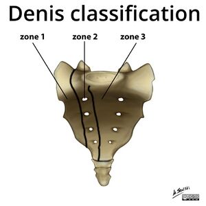
[[File:Sacrum Denis zones.jpeg|thumb|Denis classification of the zones of the sacrum]] | [[File:Sacrum Denis zones.jpeg|thumb|Denis classification of the zones of the sacrum]] | ||
{{Main|Sacrum}} | {{Main|Sacrum}} | ||
The sacrum is a triangular bone and when talking about | The sacrum is a triangular bone and when talking about the broad group of traumatic sacral fractures, the sacrum is viewed as having three zones: | ||
* Zone 1: The sacral ala lateral to the foraminae | * Zone 1: The sacral ala lateral to the foraminae. The vast majority of sacral insufficiency fractures occur in this zone. | ||
* Zone 2: the area involving the neural foraminae | * Zone 2: the area involving the neural foraminae | ||
* Zone 3: medial the the neural foraminae, containing the sacral bodies. | * Zone 3: medial the the neural foraminae, containing the sacral bodies. | ||
== Pathology == | == Pathology == | ||
Insufficiency fractures are a subtype of stress fracture where normal stress is applied to bone with reduced elastic resistance. This is usually due to osteoporosis. Other causes of insufficiency fractures are metastatic disease and bone marrow infiltration processes. | Insufficiency fractures are a subtype of stress fracture where normal stress is applied to bone with reduced elastic resistance. This is usually due to osteoporosis, which is usually primary. Other causes of insufficiency fractures are metastatic disease and bone marrow infiltration processes. | ||
They most commonly affect the sacral ala, in a sagittal line lateral to the sacral foraminae and medial to the sacral iliac joints, called zone 1. There is an equal prevalence of unilateral versus bilateral fractures. In some cases there is a fracture line component in the axial plane. | They most commonly affect the sacral ala, in a sagittal line lateral to the sacral foraminae and medial to the sacral iliac joints, called zone 1. There is an equal prevalence of unilateral versus bilateral fractures. In some cases there is a fracture line component in the axial plane. | ||
The reason that the sacral ala are affected is because in osteoporosis there isn't uniform bone loss. There is relatively increased loss of bony trabeculae in the sacral alae compared to the vertebra bodies. | The reason that the sacral ala are affected is because in osteoporosis there isn't uniform bone loss. There is relatively increased loss of bony trabeculae in the sacral alae compared to the vertebra bodies.<ref name=":0" /> | ||
There | There are concomitant pubic rami fractures in 78% of affected individuals.<ref name=":1">{{Cite journal|last=Tsiridis|first=E.|last2=Upadhyay|first2=N.|last3=Giannoudis|first3=P. V.|date=2006-12|title=Sacral insufficiency fractures: current concepts of management|url=https://pubmed.ncbi.nlm.nih.gov/16855863|journal=Osteoporosis international: a journal established as result of cooperation between the European Foundation for Osteoporosis and the National Osteoporosis Foundation of the USA|volume=17|issue=12|pages=1716–1725|doi=10.1007/s00198-006-0175-1|issn=0937-941X|pmid=16855863}}</ref> There can also be additional fractures in the superior acetabulum and iliac wing. It is thought that the sacral ala fractures happen first, about 3-4 months prior to the pubic rami fractures. It is thought that the sacral ala fractures results in a cascade of abnormal biomechanics with disrupted in one area leading to increased stress elsewhere. The follow on pubic rami fractures more commonly have a prolonged course of healing.<ref name=":0" /><ref name=":1" /> | ||
== Clinical Features == | == Clinical Features == | ||
=== History === | === History === | ||
In two thirds of patients there is no trauma, and when there is trauma it is usually minor. Patients will commonly have diffuse low back pain with radiation to the buttock, hip, or groin. 45% of patients have a history of malignancy.<ref name=":0" /> | In two thirds of patients there is no trauma, and when there is trauma it is usually minor. Patients will commonly have diffuse low back pain with radiation to the buttock, hip, or groin.<ref name=":0" /> The pain is usually exacerbated by weight bearing.<ref name=":1" /> 45% of patients have a history of malignancy.<ref name=":0" /> | ||
=== Examination === | === Examination === | ||
There may be lumbosacral spine tenderness. There is usually no neurological deficit, however rarely patients can manifest with a [[Lumbar Radicular Pain and Radiculopathy|sacral radiculopathy]] or even [[Cauda Equina Syndrome|cauda equina syndrome]]. | Gait is usually antalgic. There may be lumbosacral spine tenderness and pain over the lateral aspect of the sacrum. There is pubic rami tenderness in the presence of associated pubic rami fractures. Sacroiliac provocation tests are often positive. There is usually no neurological deficit, however rarely patients can manifest with a [[Lumbar Radicular Pain and Radiculopathy|sacral radiculopathy]] or even [[Cauda Equina Syndrome|cauda equina syndrome]].<ref name=":0" /><ref name=":1" /> | ||
== Investigations == | == Investigations == | ||
=== Blood Tests === | |||
ALP may be slightly raised and can be useful if plain films are normal and high tech imaging can't be easily accepted. After diagnosis on imaging secondary causes of osteoporosis may need to be tested for: TSH, PTH, calcium, phosphorus, albumin, vitamin D, urinary calcium, creatinine, FBC, LFTs, CRP, ESR, and SPE.<ref name=":1" /> | |||
=== Plan Films === | === Plan Films === | ||
Plain films are not very sensitive at picking up sacral insufficiency fractures, with them only being detected in 20-38% of cases.. Part of this is because dedicated sacral views are often not taken. When visible there is a vertical sclerotic band in zone 1 in 57% of cases, with a fracture line in 12% of cases.<ref name=":0" /> | Plain films are not very sensitive at picking up sacral insufficiency fractures, with them only being detected in 20-38% of cases.. Part of this is because dedicated sacral views are often not taken. The correct views are AP and lateral views of the pelvis plus/minus inlet and outlet views. When visible there is a vertical sclerotic band in zone 1 in 57% of cases, with a fracture line in 12% of cases.<ref name=":0" /> | ||
=== High Tech Imaging === | === High Tech Imaging === | ||
| Line 41: | Line 44: | ||
=== DEXA === | === DEXA === | ||
Almost all patients will have severe osteopenia on DEXA imaging. | Almost all patients will have severe osteopenia on DEXA imaging which is the gold standard for measuring bone mineral density. | ||
== Management == | == Management == | ||
The optimal treatment approach is unknown. Most patients improve with conservative care. It varies between 6 to 15 months for recovery. However 50% do not return to their premorbid state, and mortality is 14.3%.<ref name=":0" /> | The optimal treatment approach is unknown. Most patients improve with conservative care. It varies between 6 to 15 months for recovery. However 50% do not return to their premorbid state, and mortality is 14.3%.<ref name=":0" /> | ||
Prolonged bedrest | Prolonged bedrest is sometimes recommended but this carries the potential for increased morbidity. For example it is important to be alert to venous thromboembolism. DVTs occur in 29-61% of cases, and PE in 2-12%. Other complications of bedrest in elderly include sarcopenia, infection, and a multitude of other potential problems. Mobilisation can encourage bone growth. Therefore it is probably best to mobilise within the limits of pain tolerance. Walking aids such as walking frames can be helpful to encourage gradual mobilisation. Hydrotherapy may be helpful. | ||
Usually antiresorptive medication (bisphosphanates are first line in New Zealand as of time of writing in 2022) and vitamin D are prescribed for preventative care. | |||
== References == | == References == | ||
[[Category:Fractures]] | [[Category:Fractures]] | ||
[[Category:Pelvis, Hip and Thigh Conditions]] | [[Category:Pelvis, Hip and Thigh Conditions]] | ||
Revision as of 19:35, 15 November 2022
Epidemiology
This condition classical affects osteoporotic elderly women. The mean age is 70-75, with almost all patients older than 55.[1]
Other risk factors include prior pelvic radiation (prevalence 21-89%), prolonged glucocorticoid use, rheumatoid arthritis, multiple myeloma, Paget disease, renal osteodystrophy, and hyperparathyroidism. [1]
Anatomy
- Main article: Sacrum
The sacrum is a triangular bone and when talking about the broad group of traumatic sacral fractures, the sacrum is viewed as having three zones:
- Zone 1: The sacral ala lateral to the foraminae. The vast majority of sacral insufficiency fractures occur in this zone.
- Zone 2: the area involving the neural foraminae
- Zone 3: medial the the neural foraminae, containing the sacral bodies.
Pathology
Insufficiency fractures are a subtype of stress fracture where normal stress is applied to bone with reduced elastic resistance. This is usually due to osteoporosis, which is usually primary. Other causes of insufficiency fractures are metastatic disease and bone marrow infiltration processes.
They most commonly affect the sacral ala, in a sagittal line lateral to the sacral foraminae and medial to the sacral iliac joints, called zone 1. There is an equal prevalence of unilateral versus bilateral fractures. In some cases there is a fracture line component in the axial plane.
The reason that the sacral ala are affected is because in osteoporosis there isn't uniform bone loss. There is relatively increased loss of bony trabeculae in the sacral alae compared to the vertebra bodies.[1]
There are concomitant pubic rami fractures in 78% of affected individuals.[2] There can also be additional fractures in the superior acetabulum and iliac wing. It is thought that the sacral ala fractures happen first, about 3-4 months prior to the pubic rami fractures. It is thought that the sacral ala fractures results in a cascade of abnormal biomechanics with disrupted in one area leading to increased stress elsewhere. The follow on pubic rami fractures more commonly have a prolonged course of healing.[1][2]
Clinical Features
History
In two thirds of patients there is no trauma, and when there is trauma it is usually minor. Patients will commonly have diffuse low back pain with radiation to the buttock, hip, or groin.[1] The pain is usually exacerbated by weight bearing.[2] 45% of patients have a history of malignancy.[1]
Examination
Gait is usually antalgic. There may be lumbosacral spine tenderness and pain over the lateral aspect of the sacrum. There is pubic rami tenderness in the presence of associated pubic rami fractures. Sacroiliac provocation tests are often positive. There is usually no neurological deficit, however rarely patients can manifest with a sacral radiculopathy or even cauda equina syndrome.[1][2]
Investigations
Blood Tests
ALP may be slightly raised and can be useful if plain films are normal and high tech imaging can't be easily accepted. After diagnosis on imaging secondary causes of osteoporosis may need to be tested for: TSH, PTH, calcium, phosphorus, albumin, vitamin D, urinary calcium, creatinine, FBC, LFTs, CRP, ESR, and SPE.[2]
Plan Films
Plain films are not very sensitive at picking up sacral insufficiency fractures, with them only being detected in 20-38% of cases.. Part of this is because dedicated sacral views are often not taken. The correct views are AP and lateral views of the pelvis plus/minus inlet and outlet views. When visible there is a vertical sclerotic band in zone 1 in 57% of cases, with a fracture line in 12% of cases.[1]
High Tech Imaging
MRI has a sensitivity of close to 100%. It is very important that coronal oblique views of the sacrum are included. The fracture may be missed if "lumbar spine MRI" is requested. In New Zealand lumbar spine MRI and pelvis MRI are two separate scans for funding and insurance purposes.[1]
CT has a sensitivity of 60-75%, however it can be helpful for surgical planning as well as differentiating between metastatic disease and fracture.[1]
DEXA
Almost all patients will have severe osteopenia on DEXA imaging which is the gold standard for measuring bone mineral density.
Management
The optimal treatment approach is unknown. Most patients improve with conservative care. It varies between 6 to 15 months for recovery. However 50% do not return to their premorbid state, and mortality is 14.3%.[1]
Prolonged bedrest is sometimes recommended but this carries the potential for increased morbidity. For example it is important to be alert to venous thromboembolism. DVTs occur in 29-61% of cases, and PE in 2-12%. Other complications of bedrest in elderly include sarcopenia, infection, and a multitude of other potential problems. Mobilisation can encourage bone growth. Therefore it is probably best to mobilise within the limits of pain tolerance. Walking aids such as walking frames can be helpful to encourage gradual mobilisation. Hydrotherapy may be helpful.
Usually antiresorptive medication (bisphosphanates are first line in New Zealand as of time of writing in 2022) and vitamin D are prescribed for preventative care.
References
- ↑ 1.00 1.01 1.02 1.03 1.04 1.05 1.06 1.07 1.08 1.09 1.10 Lyders, E. M.; Whitlow, C. T.; Baker, M. D.; Morris, P. P. (2010-02). "Imaging and treatment of sacral insufficiency fractures". AJNR. American journal of neuroradiology. 31 (2): 201–210. doi:10.3174/ajnr.A1666. ISSN 1936-959X. PMC 7964142. PMID 19762463. Check date values in:
|date=(help) - ↑ 2.0 2.1 2.2 2.3 2.4 Tsiridis, E.; Upadhyay, N.; Giannoudis, P. V. (2006-12). "Sacral insufficiency fractures: current concepts of management". Osteoporosis international: a journal established as result of cooperation between the European Foundation for Osteoporosis and the National Osteoporosis Foundation of the USA. 17 (12): 1716–1725. doi:10.1007/s00198-006-0175-1. ISSN 0937-941X. PMID 16855863. Check date values in:
|date=(help)


