Shoulder Biomechanics
The shoulder girdle links the upper extremity to the trunk. Working with the elbow, it allows the hand to be positioned in space. It needs a high level of structural protection and functional control. The biomechanics of the shoulder are quite complex. Movement involves a complex interaction of static and dynamic stabilisers and free and coordinated motion between all four joints.
Shoulder Complex Structure
The shoulder complex (shoulder girdle) consists of four joints: glenohumeral (GHJ), acromioclavicular (ACJ), sternoclavicular (SCJ), scapulothoracic joint (STJ). Some individuals also have a coracoclavicular joint (CCJ). These joints need to have free motion and coordinated action, and motion occurs in all of them with any significant arm movement.
Sternoclavicular Joint
- Main article: Sternoclavicular Joint
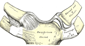
The SCJ is formed by the articulations of the proximal clavicle with the clavicular notch of the manubrium and with the cartilage of the first rib. It is a saddle shaped joint with a fibrocartilaginous disc.
Stability of the SCJ is provided by very strong ligaments, including the sternoclavicular, costoclavicular, and interclavicular ligaments, as well as by the disc, capsule, and muscular attachments. The costoclavicular ligament provides the principal support.
The SCJ provides the major axis of rotation for motion of the clavicle and scapula. The clavicle is able to move in three degrees of freedom.
- Elevation/Depression: Elevation refers to superior clavicle movement, and depression refers to inferior clavicle movement. This occurs between the clavicle and the meniscus. The range of motion is approximately 30 to 40 degrees.
- Protraction/Retraction: Protraction refers to anterior movement of the clavicle, and retraction refers to posterior movement of the clavicle. This occurs between the manubrium and the meniscus. The range of motion is approximately 30 to 35 degrees
- Rotation: The clavicle can rotate anteriorly and posteriorly along its long axis. The range of motion is approximately 40 to 50 degrees.
The close-packed position is with maximal shoulder elevation.
Acromioclavicular Joint
- Main article: Acromioclavicular Joint
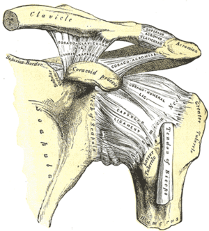
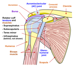
The ACJ if formed by the articulation of the acromion process of the scapula with the distal clavicle. It is an irregular diarthrodial joint and frequently has a fibrocartilaginous disc. Stability is provided by the coracoclavicular ligaments. There are many variations of the ACJ.
It is able to move in all three planes. Rotation occurs at the ACJ during arm elevation. The close-packed position is with abduction of the humerus to 90 degrees.
Coracoclavicular Joint
This is an anomalous joint formed at the junction of the coracoid process of the scapula and the inferior surface of the clavicle. They are connected by the coracoclavicular ligament. The prevalence is approximately 10%. Some texts describe this structure as normally being a syndesmosis that doesn't permit much movement.
Glenohumeral Joint
- Main article: Glenohumeral Joint Anatomy
The GHJ is the synovial ball-and-socket articulation between the humeral head and the glenoid fossa of the scapula. The GHJ is the most dynamic and mobile joint in the body.
The head of the GHJ is almost hemispherical and only 25-30% of the surface is in contact with the glenoid fossa. The glenoid fossa also has a less curved shape compared to the humeral head, and this allows for sliding movements.
The shoulder joint is reliant on ligamentous and muscular structures for stability. Stability is provided by static and dynamic components. These components restrain the humeral head and keep it in the glenoid fossa. The close-packed position is with the humerus abducted and externally rotated.
Passive Stabilisers
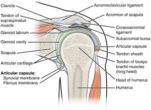
The passive static stabilisers include the articular surface, glenoid labrum, joint capsule, and ligaments. The articular cartilage is thicker peripherally and the joint is fully sealed providing negative pressure to resist dislocation at low forces.
The glenoid labrum is a dense fibrous and fibrocartilagenous tissue that encircles the glenoid fossa. It receives support from the surrounding glenohumeral ligaments and biceps brachii tendon. It is triangular in cross-section. The function is to deepen the fossa and add stability to the joint. The contact area is increased to 75% and the concavity is increased by 5mm to a total of 9mm.
The joint capsule has around twice the volume of the humeral head. It is loose in mid-range and tightens in various extreme ranges. E.g. in extreme abduction and external rotation the inferior capsule tightens. In flexion and internal rotation the anterosuperior capsule is tightened.
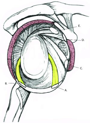
The ligaments are also part of the static stabilisers.
- Glenhohumeral ligaments: The superior, middle, and inferior glenohumeral ligaments on the anterior side are discrete extensions of the anterior glenohumeral joint capsule.
- Superior glenohumeral ligament: originates from the anterosuperior labrum, anterior to long head of biceps, inserting onto the lesser tuberosity. It is only well developed in 50% of individuals. It acts to restrain inferior translation in the adducted position
- Middle glenohumeral ligament: originates inferior to the superior glenohumeral ligament and inserts laterally on the lesser tuberosity. It is absent in 30% and has variable anatomy. It acts as a secondary restraint to inferior translation in the abduction and externally rotated position, and also restrains against anterior translation
- Inferior glenohumeral ligament: originates from the inferior aspect of the labrum and inserts on the anatomic neck of the humerus. It is the prime anterior stabiliser of the shoulder in the 90° abducted position. The anterior band is itght with abduction and external rotation, resisting anterior translating. The posterior band is tight with internal rotation and abduction resisting posterior translation. The ligament also resists inferior translation in the abducted position.
- Coracohumeral ligament: On the superior side there is the coracohumeral ligament, originating from the lateral coracoid and inserting on the anatomic neck of the humerus. The coracohumeral ligament is tight with arm adduction and resists inferior translation of the humeral head. In arm movements it resists posterior translation of the humeral and supports the weight of the arm. This ligament is more important in those with less well develop superior glenohumeral ligaments.
Dynamic Stabilisers
Dynamic stabilisation mainly occurs in mid-range. It is provided by the muscles that contract in a coordinated manner to pull the humeral head towards 1-2mm of the centre of the glenoid fossa. Prior to motion of the humerus, tension is developed in the rotator cuff and biceps tendons, increasing stability. With asymmetric contraction, the humeral head can be moved to the desired position. They also rotate and depress the humeral head during arm elevation keeping it in position.
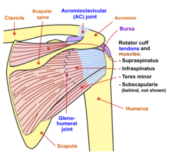
The tendons of the four rotator cuff muscles join the joint capsule and form a collagenous cuff around the GHJ surrounding the posterior, superior, and anterior sides. The posterior rotator cuff muscles provide posterior stability. The subscapularis provides anterior stability and contributes to internal rotation. The fibres of the rotator cuff muscles mingle with one another increasing their tension and functional power.
The long head of biceps brachii restrains anterior and superior humeral head translation. Deltoid and other scapulothoracic muscles position the scapula in a position to maximise stability of the GHJ.
Support Overview
Anterior shoulder joint: support is provided by the capsule, glenoid labrum, glenohumeral ligaments, capsular reinforcements, coracohumera ligament, subscapularis fibres, and pectoralis major. The muscles merge with the joint capsule. The coracohumeral and the middle glenohumeral ligaments support and hold up the relaxed arm; and support the arm through abduction, external rotation, and extension.
Posterior shoulder joint: support is provided by the capsule, glenoid labrum, and muscle fibres of teres minor and infraspinatus. The muscles also blend with the capsule.
Superior shoulder joint: support is provided by the glenoid labrum, coracohumeral ligament, overlying muscles. Supraspinatus and long head of biceps brachii reinforce the capsule. Overlying the supraspinatus is the subacromial bursa and coracoacromial ligament forming an arch underneath the ACJ.
Inferior shoulder joint: has minimal reinforcement by the capsule and long head of triceps brachii.
Scapulothoracic Joint
The STJ refers to the region between the anterior scapula and the thoracic wall. It is not a bone to bone connection but rather a physiologic joint. The scapula rests on the serratus anterior and subscapularis.
The STJ has two major functions
- Range of motion increase: It increases the total range of motion beyond the 120° generated in the GHJ. There is 1° of scapulothoracic elevation for every 2° of glenohumeral elevation.
- Large lever for scapula muscles: The size and shape of the scapula means that small muscles can provide enough torque to be effective.
Seventeen muscles attach to or originate from the scapula. They include the levator scapula, trapezius, rhomboids, serratus anterior, pectoralis minor, and subclavius. They provide the following functions:
- Scapula stabilisation: they turn the scapula into a rigid base. For example the levator scapula, trapezius, and rhomboids stabilise the shoulder against added weight when lifting.
- Movement facilitation: they position the glenohumeral joint in an appropriate position. In overhand throwing the rhomboids contract to bring the shoulder posteriorly during the preparatory phase of the throw.
The scapula moves along the thorax due to ACJ and SCJ movements. The total range of motion of the STJ is approximately 60° of motion for 180° of arm abduction or flexion. About 65% of this range occurs at the SCJ, and 35% at the ACJ. The clavicle functions as a crank on the scapula as it elevates and rotates to elevate the scapula.
The scapula has the following movements:
- Protraction or abduction, and retraction or adduction: This refers to the scapula moving anteriorly and posteriorly about a vertical axis. This occurs as the acromion moves on the meniscus and as the scapula rotates around the medial coracoclavicular ligament. The range is 30-50°.
- Upward and downward rotation: This refers to the base of the scapula swinging laterally and medially in the frontal plane. The range is around 60°
- Elevation and depression: This refers to movement of the scapula up and down. This occurs at the ACJ. The range is about 30°.
The position of the clavicle also plays a role in scapula movement. The SCJ movements are opposite to the ACJ movements for elevation, depression, protraction, and retraction; but not for rotation. E.g. with elevation at the ACJ, there is depression at the SCJ.
Bursae
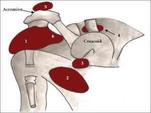
There are several bursae that reduce friction between layers of tissue and these include the subscapularis, subcoracoid, and subacromial bursae. The most well known, the subacromial bursa, sits in the subacromial space between the acromion and the coracoacromial ligament above and the glenohumeral joint below. It cushions the supraspinatus from the overlying acromion. The subscapularis and subcoracoid bursae are sometimes merged. They reduce friction of the superficial fibres of subscapularis against the neck of the scapula, the head of the humerus, and the coracoid process.
Movements of the Shoulder Complex
Range of Motion
The shoulder can move in the following ranges:
- Flexion/Extension: The arm has a range of around 165° to 180° of flexion to approximately 30° to 60° of hyperextension in the sagittal plane. Flexion range is reduced with a position of external rotation; in maximal external rotation, flexion is only 30°.
- Translation: Sliding movements also occur. During passive flexion there is anterior translation, and with passive extension there is posterior translation of the humeral head on the glenoid fossa.
- Abduction/Adduction: Abduction range is 150° to 180°. Abduction is reduced by internal rotation; in maximal internal rotation, abduction is only 60°. In adduction, the arm can continue past neutral to approximately 75° of hyperadduction across the body
- Internal/External Rotation: The total range of rotation is 120° to 180° combining internal rotation of 60° and external rotation of 90°. In abduction, rotation is constrained; in 90° of abduction, rotation is only 90°
- Horizontal Flexion/Extension, Adduction/Abduction: The range is 135° of horizontal flexion or adduction and 45° of horizontal extension or abduction
Scapulohumeral Rhythm
Most commonly motion of the humerus involves some motion of all shoulder joints. However some amount of GHJ motion can occur while the others are stabilised. The scapulohumeral rhythm refers to the coordination of scapular and humeral movements with the four joints of the shoulder complex all working together. This rhythm allows a greater range of motion at the shoulder than if the scapula was a fixed structure.
In the the early stages of abduction or flexion, the movement is primarily at the GHJ. However there are also stabilising motions of the scapula.
Once past 30° of abduction or 45-60 degrees of flexion, there is 5° of humeral movement for every 4° of scapular movement on the thorax (5:4 ratio). When looking at the total range of motion from 0-180° of abduction or flexion, the full 180° is made up of 120° of GHJ motion and 60° of scapular motion, i.e. a 2:1 ratio.
There are contributing movements of the ACJ (20°), sternoclavicular joint (40°), and posterior clavicular rotation (40°). During the first 90° of arm elevation (in all planes), the clavicle is also elevated at the SCJ, and rotation occurs at the ACJ.
At 90° of abduction, the greater tuberosity approximates the acromion process with soft tissue compression limiting further abduction. With external rotation of the arm, 30° further abduction can occur because the greater tuberosity is moved out from the coracoacromial arch. On the other hand, if the arm is internally rotated, there is only 60°of abduction. Therefore external rotation occurs with abduction to about 160°. Full range of abduction requires some extension of the upper trunk
Muscle Action
The muscles that contribute to abduction and flexion are similar.
The deltoid contributes 50% of the muscular force with abduction or flexion (i.e. elevation). With increased abduction there is greater contribution from the deltoid and it is most active from 90-180°. Deltoid and supraspinatus are active throughout the range of arm elevation, but supraspinatus has a larger role in initiating abduction.
Deltoid's anterior head is a strong flexor and internal rotator, the middle head an abductor, and posterior head an extensor and external rotator.
The deltoid cannot abduct or flex the arm without humeral head stabilisation from the rotator cuff. The rotator cuff contributes about 50% of the forces for abduction and flexion. With flexion stability is provided by supraspinatus, infraspinatus and latissimus dorsi. With pure abduction there is similar activity but subscapularis provides stabilisation through eccentric contraction.
In early abduction or flexion, the deltoid is oriented vertically. Supraspinatus assists deltoid and compresses the humeral head to stop it translating superiorly from deltoid action. The rotator cuff muscles as a group all contract and compress the humeral head into the glenoid fossa. The teres minor, infraspinatus, and subscapularis pull the humerus down which stabilises it in elevation. Latissimus dorsi also eccentrically contracts with elevation.
Above 90° of flexion or abduction there is reduced rotator cuff action, except for the supraspinatus. In this range, deltoid pulls the humeral head down As noted above in this range there is external rotation. With 20° or more of external rotation, biceps brachii is also able to abduct the arm.
With arm abduction or flexion, the shoulder girdle undergoes protraction or abduction, elevation, and upward rotation with posterior clavicular rotation maintaining the glenoid fossa in an optimal position. After deltoid and teres minor initiate elevation, serratus anterior and trapezius create lateral, superior, and rotational motions of the scapula, especially from 90-180°. Serratus anterior also holds the scapula to the thoracic wall by preventing the medial border from lifting off.
With lowering of the arm, i.e. adduction or extension, there is eccentric muscular action and retraction, depression, and downward rotation of the shoulder girdle along with forward clavicular rotation. Rhomboid, teres major, and latissimus dorsi work to control the arm during lowering. Pectoralis minor depresses and downwardly rotates the scapula, while the middle and lower portions of trapezius retract the scapula along with rhomboid.
If the arm is forcefully lowered or lowered against resistance then there is concentric muscular action. With external resistance the latissimus dorsi, teres major, and sternal portion of pectoralis major assist provide concentric action.
External rotation is produced by infraspinatus, posterior head of deltoid, and teres minor muscles. The prime external rotator is infraspinatus. Subscapularis is also active to prevent anterior displacement of the humeral head.
Internal rotation is produced by subscapularis, latissimus dorsi, teres major, and sternal head of pectoralis major. Subscapularis is active during all phases but decreased activity with extremes of abduction. Pectoralis major and latissimus dorsi also decrease with increased abduction, while the posterior and middle heads of deltoid contrict eccentrically to assist.
Extension is achieved through action from the posterior and middle heads of deltoid. Supraspinatus and subscapularis are also active throughout extension with eccentric activity preventing anterior dislocation.
Muscular Strength
Concentric adduction strength is twice that of abduction strength. Extension is the next strongest and this is slightly stronger than flexion. Abduction is the third strongest. In other words, with lowering of the arm involving extension and adduction there is greater strength than during raising of the arm involving flexion and abduction.
With isometric contraction however, the flexors are stronger than the extensors.
Rotational strength is the weakest joint force, and internal rotation is stronger than external rotation.
Summary
- The sternoclavicular joint, which connects the medial end of the clavicle to the manubrium, links the upper extremity to the thorax. The articular disc within the joint and the ligaments that surround it provide stability while allowing for significant rotation of the clavicle. Little relative motion is seen between the clavicle and acromion at the acromioclavicular joint.
- The glenohumeral joint, while known to be a precise articulation, is inherently unstable because the glenoid fossa is shallow and is able to contain only approximately one third of the diameter of the humeral head. Stability is instead provided by the capsular, muscular, and ligamentous structures that surround it.
- The three glenohumeral ligaments (superior, middle, and inferior) are discrete extensions of the anterior glenohumeral joint capsule and are critical to shoulder stability and function.
- The inferior glenohumeral ligament has the most functional significance (particularly the anterior band), acting as the primary anterior stabilizer of the shoulder when the arm is abducted 90°.
- The integrity of the shoulder capsule and the negative intra-articular force it maintains also plays an integral part in maintaining shoulder stability.
- Elevation of the arm involves motion at both the glenohumeral joint and the scapulothoracic articulation (2:1).
- Movements of the spine assist the shoulder in positioning the upper extremity in space.
- The muscles around the shoulder contribute to stability via a barrier effect by producing compressive forces on the glenohumeral joint and by eccentric contraction.
- The glenohumeral joint is a major load-bearing joint with forces equivalent to one-half body weight produced when holding the arm in an outstretched position.
References
Nordin, Margareta, and Victor H. Frankel. Basic biomechanics of the musculoskeletal system. Philadelphia: Wolters Kluwer Health/Lippincott Williams & Wilkins, 2012.
