Shoulder Joint Injection
| Shoulder Joint Injection | |
|---|---|
| Indication | Adhesive Capsulitis |
| Syringe | 5-10mL |
| Needle | 21g 40-50mm |
| Steroid | 40mg triamcinolone |
| Local | 4mL 1% |
| Volume | 5mL (11mL if hydrodilating and aspirate back) |
Glenohumeral joint injections (often referred to as shoulder injections ) are performed as part of a number of therapeutic and imaging procedures using a variety of approaches and modalities. The underlying principles shared by all techniques are to avoid damage to the glenoid labrum, long head of biceps tendon, surrounding neurovascular structures and articular cartilage.
Anatomy
- See also: Shoulder_Biomechanics#Glenohumeral_Joint
The glenohumeral joint is a synovial diarthrodial joint where the humeral head articulates with the glenoid fossa of the scapula. The joint has static and dynamic stabilisers.
A normal joint will usually have a capacity of 8-15 mL. This will be reduced in adhesive capsulitis.
Indications
Injection into the glenohumeral joint may be necessary in the following settings:
- diagnostic and/or therapeutic corticosteroid +/- local anaesthetic injection
- glenohumeral (shoulder) arthrography
- glenohumeral (shoulder) hydrodilatation
Contraindications
Anticoagulation is a relative contraindication and should be assessed in the context of the risks of ceasing anticoagulation versus the risk of haemarthrosis. It some settings it will be best to avoid arthrography entirely or consider using indirect arthrography.
Pre-procedural Evaluation
Routine patient interactions are carried out (the procedure is explained to the patient, informed consent obtained, allergy and comorbidity history obtained, time-out performed including ensuring the correct side is being investigated, etc).
The shoulder needs to be exposed and skin examined for active infection.
Equipment
- Sterile procedure pack, wash, gloves and gown
- Local anaesthetic for skin (e.g. 1%/2% lignocaine) with needle (e.g. 23 or 25 G needle) and syringe
- Needle: 21 or 25-gauge spinal needle, 2 to 3.5 inch in length depending on body habitus.
- Syringe for injectable with injectate: e.g. 4-9mL of local anaesthetic and 1mL of corticosteroid.
- Syringe for contrast if doing it under fluoroscopy
- Short connecting tube (optional)
- Medium to high frequency transducer, curvilinear is often preferred, if doing it under ultrasound guidance.
- Dressing
Technique
A variety of approaches, both anterior and posterior, have been described to inject the glenohumeral joint. The most common approaches are landmark, ultrasound, and fluoroscopy.
An RCT found that ultrasound-guided injections where more clinically effective and more cost-effective than nonguided injections.[1]
Ultrasound Guided
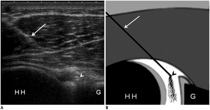
Both anterior and posterior approaches (see landmark for reference) can be performed under ultrasound guidance. Injection has been described through the rotator cuff interval.[2]
Posterior Approach
The following is a posterior approach described by Malanga and colleagues.[3]
- Position: Lateral recumbent with the affected side facing up. The arm at the patients side in neutral rotation which can be bolstered under the arm for patient comfort.
- Transducer position: Anatomic axial oblique plane, parallel to the fibres of the infraspinatus tendon, over the posterior aspect of the glenohumeral joint
- Needle orientation: in-plane to probe. The bevel should face the articular surface of the humerus so that the articular cartilage is not gouged.
- Needle approach: Posterior lateral to anterior medial. Needle visualised entering into the joint space between the humeral head and glenoid labrum.
- Target: Posterior glenohumeral joint, with the needle tip between the humeral head and glenoid labrum
- Visualise: Injectate flowing into the joint space.
You can use the anaesthetising needle to determine the exact needle trajectory before using the spinal needle for intraarticular injection. The needle needs to enter at quite a steep angle, and so avoid shallow initial placement.
Take care to avoid needle placement superficial and medial to the glenohumeral joint which raises risk of injury ot the neurovascular structures in the spinoglenoid notch.
If hydrodilating inject for example for a total volume of 11mL, use half bupivacaine and half saline intraarticularly. Then wait a few seconds and aspirate as much as possible back. Then inject steroid.
You can also do the procedure sitting with the patients affected arm adducted to open up the posterior joint space.
Fluoroscopy Guided
Regardless of technique meticulous sterile technique and generous antiseptic prep to the skin should be applied.
- Anterior approach
The patient is placed supine with the arm somewhat externally rotated (palm facing upwards). Note, excessive external rotation not only may be painful, it will also tighten the anterior capsule reducing the space anteriorly.
Skin entry is marked over the upper medial quadrant of the humeral head. This is the rotator cuff interval, avoiding the tendons of supraspinatus, subscapularis and biceps tendon. Alternatively, a location somewhat lower down along the humeral head can be chosen, requiring passing through the subscapularis tendon 5,6. This notwithstanding, what is critical is that the needle is lateral to the medial humeral articular edge to avoid damaging the glenoid labrum.
The needle is then introduced vertically (needle tip overlying the hub) along the axis of the x-ray beam at the marked site until articular cartilage is encountered.
Intra-articular position is confirmed by the introduction of a small amount of contrast that should be seen to outline the joint space and the subcoracoid recess
- Posterior approach
The patient is placed prone with the shoulder to be injected elevated. Imaging is then orientated to see the joint line tangentially (i.e. joint space is visualised without overlap of glenoid and humeral head)
Skin entry is marked over the inferomedial aspect of the articular surface, superomedial to the anatomical neck of the humerus (the site of capsular attachment).
The needle is then introduced vertically along the axis of the x-ray beam at the marked site until articular cartilage is encountered
Intra-articular position is confirmed by the introduction of a small amount of contrast
Landmark Guided
- Posterior Approach
- Patient position: sitting with arms folded, thus opening up the posterior joint space
- Identify posterior angle of acromion with thumb, and coracoid process with index finger
- Insert needle directly below angle and pass anteriorly obliquely towards coracoid process until needle gently touches intra-articular cartilage
- Injection solution as a bolus
- Anterior Approach
- Patient position: arm is held is slight lateral rotation
- The needle is inserted on the anterior surface between the coracoid process and the lesser tuberosity of the humerus, aimed posteromedially towards the spine of the scapula
- This is less preferable to the posterior approach due to patient visualisation, sensitive flexor skin surfaces, and more neurovascular structures present.
Complications
Extracapsular injection is probably the most common complication. The most serious complication is septic arthritis. Haemarthrosis is also rarely encountered.
Aftercare
Videos
References
Part or all of this article or section is derived from Glenohumeral joint injection (technique) by Andrew Murphy and Assoc Prof Frank Gaillard et al., used under CC BY-NC-SA 3.0
- ↑ Sibbitt WL Jr, Band PA, Chavez-Chiang NR, Delea SL, Norton HE, Bankhurst AD. A randomized controlled trial of the cost-effectiveness of ultrasound-guided intraarticular injection of inflammatory arthritis. J Rheumatol. 2011 Feb;38(2):252-63. doi: 10.3899/jrheum.100866. Epub 2010 Nov 15. PMID: 21078710.
- ↑ Souza PM, Aguiar RO, Marchiori E, Bardoe SA. Arthrography of the shoulder: a modified ultrasound guided technique of joint injection at the rotator interval. Eur J Radiol. 2010 Jun;74(3):e29-32. doi: 10.1016/j.ejrad.2009.03.020. Epub 2009 Apr 25. PMID: 19394776.
- ↑ Malanga, Gerard A., and Kenneth R. Mautner. Atlas of ultrasound-guided musculoskeletal injections. New York: McGraw-Hill Education Medical, 2014.
Literature Review
- Reviews from the last 7 years: review articles, free review articles, systematic reviews, meta-analyses, NCBI Bookshelf
- Articles from all years: PubMed search, Google Scholar search.
- TRIP Database: clinical publications about evidence-based medicine.
- Other Wikis: Radiopaedia, Wikipedia Search, Wikipedia I Feel Lucky, Orthobullets,


