Slipping Rib Syndrome
Slipped rib syndrome is a poorly understood, but potentially under-recognised cause for chronic chest wall pain.
Anatomy
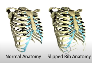
The 8th to 10th "false" rib cartilage tips normally merge inot a common arch joining to the sternum. The false ribs "piggyback" on the 7th true rib cartilage. The interchondral joints, which join the false ribs, provide some stability and flexibility with movmenet and breathing. The diaphragm connects to the false ribs, using them as an anchor point for activation.[1]
Epidemiology
The diagnosis has been documented in 1% of all general medical referrals, and 5% of new gastroenterology referrals.[1] In research by Hansen et al, the mean age was 51, 90% were female, 38% have a history of trauma, the average duration of pain was 38 months. The patients tended to be at the end of the line with 41% had suicidal ideations due to pain severity.[2]
Aetiology

In slipped rib syndrome, one or more false ribs separate, becoming "slipped" or floating. This can be due to trauma, degenerative change, and hypermobile connective tissue disorders. The condition is a joint dislocation. It most commonly involves the 10th rib, but can involve additional ribs. In Dr Adam Hansen's experience, the 10th rib is always involved.[1] Typically the affected rib will slip towards and under the rib above, impinging the intercostal nerve between the two. This can occur with simple every day movements, and can cause chronic pain.
Clinical Manifestations
The pain is dermatomal, and typically extends to the back over the costovertebral joint. It can also refer to the upper abdomen. It can be confused with visceral pathology.[1]
Assessment
Physical examination
Dr Hansen uses the following criteria for diagnosis on physical examination[1]
- Palpable separation of ≥1cm at insertion of any 8-10th rib onto the costal arch
- Same rib hypermobile on palpation
- Palpation at the separation point reproduces the patient's pain
Imaging
The costal cartilage defect is not visible on imaging. The condition can be assessed via dynamic ultrasound.[3][4] However this type of imaging assessment does not seem to be taught to MSK utlrasonographers in New Zealand at the time of writing, and so the study quality is likely to be variable.
Treatment
Nonsurgical therapies have been reported by many authors to be ineffective.[1]
Injections
Intercostal nerve blocks
Ablation
Surgical
- Rib excision
- A new technique known as Hansen repair has been described. [2] See a video of the technique here, and a webinar by Dr Hansen here.
Videos
References
- ↑ 1.0 1.1 1.2 1.3 1.4 1.5 1.6 https://www.youtube.com/watch?v=EZDFe3fC6ck
- ↑ 2.0 2.1 Hansen et al.. Minimally Invasive Repair of Adult Slipped Rib Syndrome Without Costal Cartilage Excision. The Annals of thoracic surgery 2020. 110:1030-1035. PMID: 32330472. DOI.
- ↑ Meuwly et al.. Slipping rib syndrome: a place for sonography in the diagnosis of a frequently overlooked cause of abdominal or low thoracic pain. Journal of ultrasound in medicine : official journal of the American Institute of Ultrasound in Medicine 2002. 21:339-43. PMID: 11883545. DOI.
- ↑ Van Tassel et al.. Dynamic ultrasound in the evaluation of patients with suspected slipping rib syndrome. Skeletal radiology 2019. 48:741-751. PMID: 30612161. DOI.

