Sternoclavicular Joint Pain and Instability: Difference between revisions
No edit summary |
|||
| (25 intermediate revisions by the same user not shown) | |||
| Line 1: | Line 1: | ||
{{ | {{partial}} | ||
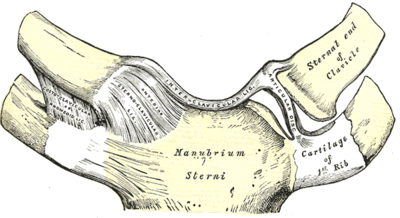
[[File:SCJ Gray.png|thumb|right|400px|The sternoclavicular joint]] | |||
This article reviews sternoclavicular instability and pain. See [[Sternoclavicular Joint]] for a review of the anatomy | This article reviews the classification, assessment, and management of sternoclavicular instability and pain. See [[Sternoclavicular Joint Anatomy]] for a review of the anatomy | ||
==Classification of Instability== | ==Classification of Instability== | ||
SCJ instability can be structure or non-structural, and causative factors may be traumatic, atraumatic, neurological, or a combination. Other than an acute first time event, instability may be persistent or recurrent. | {{See also|Sternoclavicular Joint Anatomy}} | ||
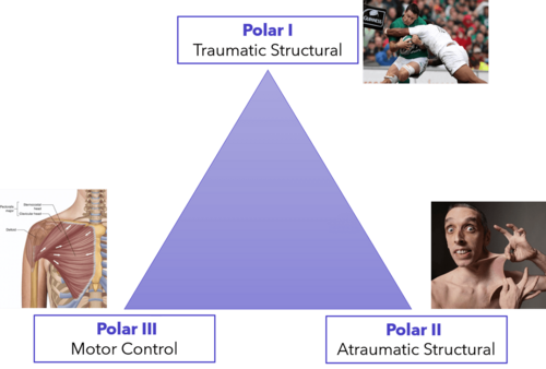
[[File:Stanmore triangle SCJ.png|500px|thumb|right|Stanmore's triangle system applied to the sternoclavicular joint]] | |||
The [[Sternoclavicular Joint Anatomy|Sternoclavicular joint (SCJ)]] is inherently unstable. The main stabilisers are the strong extrinsic ligaments, and to a lesser extent the muscular envelope. SCJ instability can be structure or non-structural, and causative factors may be traumatic, atraumatic, neurological, or a combination. Other than an acute first time event, instability may be persistent or recurrent. Dislocations are usually anterior, but can uncommonly be posterior which is more serious. | |||
Stanmore's triangle system, traditionally used for describing glenohumeral joint instability, has also been used to describe SCJ instability. There are three factors that are represented as points in a triangle: type I traumatic structural, type II atraumatic structure, and type III muscle patterning (neuromuscular) non-structural. The groups exist as a spectrum, and patients can have features of two groups, and/or move to a different pattern. With type I, there is a history of trauma such as SCJ dislocation or medial clavicular fracture. In Type II there is a structural capsular pathology without a history of "macro"-trauma but there may be micro-traumatic change. In type III there is a pathological muscle-pattern with inappropriate activation of the pectoralis major clavicular and sternal parts. This is most noticeable with eccentric overhead activities. Muscle patterning can be difficult to detect. | Stanmore's triangle system, traditionally used for describing glenohumeral joint instability, has also been used to describe SCJ instability. There are three factors that are represented as points in a triangle: type I traumatic structural, type II atraumatic structure, and type III muscle patterning (neuromuscular) non-structural. The groups exist as a spectrum, and patients can have features of two groups, and/or move to a different pattern. With type I, there is a history of trauma such as SCJ dislocation or medial clavicular fracture. In Type II there is a structural capsular pathology without a history of "macro"-trauma but there may be micro-traumatic change. In type III there is a pathological muscle-pattern with inappropriate activation of the pectoralis major clavicular and sternal parts. This is most noticeable with eccentric overhead activities. Muscle patterning can be difficult to detect. | ||
The spectrum of pathologies affecting the SCJ can be categorised into the three Stanmore instability groups. <ref name="sewell"/> | |||
*Type I traumatic structural | |||
**Medial clavicular fracture | |||
**Physeal injury (under 25) | |||
**Dislocations (posterior, anterior) | |||
*Type II atraumatic structural | |||
**Heritable connective tissue disorders causing capsular laxity (e.g. Ehlers Danlos syndrome, Marfan syndrome) | |||
**Short clavicular shape (under 25) | |||
**Osteoarthritis | |||
**Inflammatory arthritis | |||
**Infection | |||
**Sternoclavicular hyperostosis syndrome. | |||
*Type III non-structural | |||
**Pathological activation of pectoralis major. | |||
;Characteristics of the Stanmore triangle classification of joint instability<ref name="sewell"/> | |||
{| class="wikitable" | |||
! Pathology !! Type I: Traumatic structural !! Type II: Atraumatic structural !! Type III: Muscle patterning, non-structural | |||
|- | |||
| Trauma || Yes || No || No | |||
|- | |||
| Articular surface damage || Yes (e.g. disc or chondral injuries) || Not initially, but occurs when longstanding (disc attrition) || No | |||
|- | |||
| Capsular problem || Yes || Yes || Sometimes | |||
|- | |||
| Laxity || Unilateral || Uni-/bilateral || Often bilateral | |||
|- | |||
| Muscle patterning || Normal || Normal || Abnormal pectoralis major activity | |||
|- | |||
| Treatment || Physiotherapy + often ORIF, SCJ reconstruction || Physiotherapy +/- SCJ reconstruction || Physiotherapy (biofeedback) | |||
|} | |||
==Clinical Assessment== | |||
{{Red flags| | |||
*High energy injury (concomitant injuries) | |||
*Stridor, dysphagia, shortness of breath (oesophageal or tracheal compression) | |||
*Venous congestion of the face and upper limb (subclavian vein compression) | |||
*Unequal pulses (subclavian artery compression) | |||
*Subtle findings (posterior dislocation) | |||
*Red, hot, painful swollen joint (septic arthritis) | |||
*Under 25 years old (medial clavicular physeal or metaphyseal fracture-separation)}} | |||
Posterior SCJ dislocations are rare, however 30% have associated tracheal, oesophageal, or neurovascular compression, and a 3-4% mortality rate. Therefore careful assessment is required. | |||
Medial clavicular fractures and first-time traumatic SCJ dislocations are caused by direct or indirect injury. Posterior dislocation of the SCJ (referring to the medial clavicle dislocating posteriorly) can be sustained by a direct blow to the anteromedial aspect of the clavicle. Anterior displacement tends to occur through a characteristic oblique shearing extracapsular fracture in the medial clavicle (disrupting the anterior capsule +/- coraco-clavicular ligament (CCL), while sparing the posterior capsule), and can be sustained by a compressive force applied to the lateral aspect of the shoulder girdle such as when being struck from the side. With these high-energy injuries there should be a high index of suspicion for additional internal thoracic and upper limb injuries. This type of force can also cause anterior dislocation, and can be difficult to differentiate from a medial clavicular fracture. A fractured medial clavicle has a blunt pointed shape and moves obliquely in front of the sternum, while with a dislocated SCJ the medial clavicle moves forward (not across the sternum) and looks like a lump. A compressive lateral force with the arm flexed at the shoulder is another mechanism for posterior SCJ dislocation. | |||
Anterior dislocations are generally quite obvious presenting as a prominent anterior lump. Posterior dislocations on the other hand are often subtle. Pain and swelling are often localised to the joint. The patient may hold their arm across their chest to minimise both glenohumeral and scapulothoracic movement. There may be bruising below the clavicle. If there is a lack of resistance where the CCL should be, then this could suggest a fracture with incomplete CCL rupture. With posterior dilocation the scapula may be protracted and the entire shoulder girdle may be shorted and asymmetrical, with little if any visible swelling. Tracheal and/or oesophageal compression can occur with the patient having stridor, dysphagia, or shortness of breath. This requires emergent joint reduction which can be achieved in a closed method if acute, but if persistent then open surgery may be required due to distorted adherent vessel tissue behind the joint due to inflammatory changes. | |||
Compression and thrombosis of the subclavian vein can occur with posterior dislocation. In this situation the patient may have venous congestion of the face and upper limb. Subclavian artery compression is rare and may be suggested by unequal pulses. If suspicious then angiography may be required. Chest x-ray is required in order to exclude a pneumothorax, pneumomediastinum, and haemopneumothorax. Brachial plexopathy is possible and can occur following direct trauma, compression, or traction. These can be managed conservatively if it is a closed injury.<ref name="sewell"/> | |||
== Differential Diagnosis == | |||
{{DDX Box|ddx-text={{Sternoclavicular Joint Pain DDX}}|ddx-title=Differential Diagnosis of a Painful or Enlarged Sternoclavicular Joint}} | |||
Information from Davis et al.<ref>Davis et al. ExpertDDx Musculoskeletal. Elsevier; 2nd edition 2017</ref> | |||
==Radiological Investigations== | |||
===Radiographs=== | |||
Standard chest x-rays have a low sensitivity for detecting traumatic SCJ dislocation. <ref name="sewell"/> | |||
;Serendipity view | |||
In the cephalic-tilt or 'serendipity' view, the x-ray beam projects caudocephalically. In this view anterior dislocations are detected by a superiorly displaced medial clavicle, and posterior dislocations as an inferiorly displaced medial clavicle.<ref name="sewell"/> | |||
;Heinig view | |||
In this view the beam is directed perpendicular to the joint (i.e. oblique to the patient), while the patient is supine. This is a useful view. Dislocations are detected by looking at their relationship to the laterally projected manubrium. <ref name="sewell"/> | |||
===CT=== | |||
CT is superior to x-ray for diagnosing dislocation. It can provide high-resolution axial images, and a three dimensional reconstruction. It can identify abnormal anatomical variants that predispose the patient to instability. <ref name="sewell"/> | |||
===MRI=== | |||
MRI has a poorer resolution than CT, but can detect soft tissue injuries. Disc injury (especially in recurrent atraumatic instability), ligament disruption, and the adjacent neovascular structures can all be assessed. Contrast enhancement is often required to detect disc injuries. In patients with posterior dislocation, MR angiography can given better visualisation due to the tethering of adhesions in the retrosternal space distorting the anatomy.<ref name="sewell"/> | |||
==Management== | |||
===Anterior Dislocation=== | |||
The acute anterior dislocation (7-10 days) can be reduced by closed manipulation under conscious sedation, or in an operating room under general anaesthesia. Place the patient supine, and put a bolster between the scapulae. Place the arm in neutral flexion and abduction. Apply pressure over the medial clavicle and traction the arm. This almost always reduces the dislocation. Place the arm in a sling that maintains scapular protraction for up to four weeks. Recurrence is very common at > 50%. For patients with persistent instability, open reduction and stabilisation can be considered.<ref name="sewell"/> | |||
===Posterior Dislocation=== | |||
If the patient is over 25, then it can be assumed that the medial physis has closed, and closed reduction can be attempted within 7-10 days from injury. If more time has passed then there may be the formation of inflammatory adhesions and so open reduction is required. The most common closed technique is the abduction-traction technique. It is similar to anterior dislocations. Abduct the ipsilateral shoulder to 90° and apply traction. Then apply an extension force to the shoulder which may translate the medial clavicle anteriorly and untrap it from behind the manubrium. Another technique is applying a direct posterior force to both shoulders while both arms are held in adduction and traction. After reduction, the arm should be put in a figure of eight bandage for 6 weeks to encourage scapular retraction.<ref name="sewell"/> | |||
===Acute Ligament Sprains=== | |||
Ligament injuries are graded as type 1 (mild), type 2 (moderate), and type 3 (severe). Type 1 injuries are when the ligaments are stretched. In type 2 there is partial ligament rupture without instability. In type 2, the patient should be treated with sling immobilisation, analgesia, and progressive mobilisation. The patient may be permitted hand-behind-back and overhead activities after six weeks, and weight-bearing at three months. Type 3 injuries are when there is complete ligament rupture and instability.<ref name="sewell"/> | |||
===Medial Clavicular Fractures, Physeal and Disc Injuries=== | |||
Medial clavicular fracture are high-energy fractures. They are unstable and are associated with other injuries. The mortality rate is 20% within one month. They are often non- or minimally displaced and only rarely involve the SCJ. They can be treated non-operatively with immobilisation in a sling. If the fracture is significantly displaced then there may be risk to the mediastinal structures. With non-operative management >50% have persistent discomfort. | |||
In younger patients under 25 years, medial physeal fractures are commonly misdiagnosed as SCJ dislocations. All patients under 25 years of age should have gold standard imaging to detect occult medial physeal injury. Ct may only show a displaced medial clavicle if the patient is under 19 where ossification has not yet occurred. Orthopaedic input is recommended. | |||
If the patient has persistent pain then consider intra-articular disc injury. This is best imaged with MRI. It can be managed with [[Sternoclavicular Joint Injection|Steroid injections]] injections, physiotherapy, and surgery.<ref name="sewell"/> | |||
===Type I Instability (Traumatic Structural)=== | |||
With minimal loss of function and minimal symptoms, the patient can be managed non-operatively. [[Sternoclavicular Joint Injection|Steroid injections]] and physiotherapy can be considered. The main surgical option is resection of the medial clavicle with CCL reconstruction in patients with persistent symptoms. Persistent symptoms is thought to be due to failure to achieve medial clavicular control. The use of pin transfixion for stabilisation is absolutely contraindicated because it has potentially fatal complications<ref name="sewell"/> | |||
===Type II Instability (Atraumatic Structural)=== | |||
There are several pathologies that cause type II instability.<ref name="sewell"/> | |||
;Generalised ligamentous laxity | |||
The average age of presentation is 18 years old with equal gender distribution. There is a prominence of the medial clavicle and pain with overhead activities. Patients can be managed non-operatively with [[Sternoclavicular Joint Injection|Steroid injections]] and physiotherapy. Most patients do not have limitation of activity with non-operative management. 90% have persistent subluxation and 21% have ongoing pain. Surgery is not indicated as all patients treated surgically have a poor result. Atraumatic posterior dislocation is rare but surgery is indicated due to the risk of retrosternal compression. | |||
;Short Clavicle | |||
Clavicular growth occurs at the medial physis and so the risk of anterior subluxation reduces over time, and this problem is rare in patients over 25 years old. There is a higher torque stress on the SCJ during movement. Scapular retraction forces the medial clavicle anteriorly, and the vertical CCL fibres rotate. Potential issues may be delayed growth and asymmetrical scapula posture such as due to scoliosis, especially if the patient also has hypermobility. The more typical clavicular antecurve protects against this leverage.<ref name="sewell"/> | |||
;Degenerative Osteoarthritis | |||
This tends to occur in the inferomedial articular cartilage of the SCJ in the dominant limb of post-menopausal women. Patients may have pain and clicking in the joint, that is exacerbated by cross-body adduction. There may be localised swelling. [[Sternoclavicular Joint Injection|Steroid injections]] and physiotherapy are generally helpful. In persistent problems, the patient may be suitable for debridement, discectomy, and capsulorrhaphy. | |||
;Inflammatory Arthritis | |||
As a synovial joint, inflammatory arthritis can occur. SCJ is involved in 31% of patients with rheumatoid arthritis. It is often undiagnosed. A [[Sternoclavicular Joint Injection|Steroid injection]] can be helpful. The patients underlying condition should be optimised. Surgical management is less reliable than osteoarthritis. | |||
;Septic Arthritis | |||
This generally presents as a red, hot, painful, swollen joint. There may however just be localised cellulitis. The joint should be aspirated under ultrasound guidance. Intravenous antibiotics +/- surgical lavage is indicated. | |||
;Sternocostoclavicular hyperostosis syndrome (SCCH) | |||
This was first described in middle-aged Japanese men. In this condition there is hyperossification of the manubrium, sternum, clavicles, upper ribs, and peri-articular ligaments. Palmoplantar pustulosis is present in 60% of cases. It may be related to [[Diffuse Idiopathic Skeletal Hyperostosis|Diffuse idioopathic skeletal hyperostosis]] and ankylosing spondylitis. Diagnosis is mainly by exclusion. Histology shows chronic inflammation, and a few patients have growth proprionobacterium acnes. Treatment is symptomatic. Antibiotics may reduce pain and pustulosis. | |||
===Type III Instability (Non-structural)=== | |||
These patients have poor coordination of the musculature, not a problem with structure and strength. This is managed with physiotherapy and biofeedback. EMG have been used to objectively show inappropriate pectoralis major recruitment. Biofeedback involves re-learning correct muscle patterns and improving proprioception. If surgery is considered for a Type II instability, then muscle patterning should first be identified and addressed before proceeding. | |||
==Summary== | |||
*Instability can be due to traumatic structural, atraumatic structural, and/or muscle patterning pathology. | |||
*Traumatic structural instability requires careful investigation and frequently surgical intervention. | |||
*Atraumatic structural instability is more common and in most patients can be successfully managed non-operatively. Surgery has a low rate of patient satisfaction | |||
*Muscle patterning should be addressed before considering surgical intervention. | |||
==Resources== | ==Resources== | ||
See Garcia et al for an open access review on sternoclavicular joint instability. | See Garcia et al for an open access review on sternoclavicular joint instability.{{#pmid:32801951|garcia}} Also an older open access review by Sewell et al.{{#pmid:23723264|sewell}} | ||
==See Also== | |||
*[[Sternoclavicular Joint Injection]] | |||
*[[Sternoclavicular Joint Anatomy]] | |||
==References== | ==References== | ||
<references/> | |||
{{Reliable sources|synonym1="sternoclavicular joint pain"|synonym2="sternoclavicular joint instability|synonym3="sternoclavicular joint"}} | |||
[[Category:Shoulder]] | [[Category:Shoulder Conditions]] | ||
[[Category: | [[Category:Presenting Complaints]] | ||
Revision as of 19:13, 23 April 2022
This article reviews the classification, assessment, and management of sternoclavicular instability and pain. See Sternoclavicular Joint Anatomy for a review of the anatomy
Classification of Instability
- See also: Sternoclavicular Joint Anatomy
The Sternoclavicular joint (SCJ) is inherently unstable. The main stabilisers are the strong extrinsic ligaments, and to a lesser extent the muscular envelope. SCJ instability can be structure or non-structural, and causative factors may be traumatic, atraumatic, neurological, or a combination. Other than an acute first time event, instability may be persistent or recurrent. Dislocations are usually anterior, but can uncommonly be posterior which is more serious.
Stanmore's triangle system, traditionally used for describing glenohumeral joint instability, has also been used to describe SCJ instability. There are three factors that are represented as points in a triangle: type I traumatic structural, type II atraumatic structure, and type III muscle patterning (neuromuscular) non-structural. The groups exist as a spectrum, and patients can have features of two groups, and/or move to a different pattern. With type I, there is a history of trauma such as SCJ dislocation or medial clavicular fracture. In Type II there is a structural capsular pathology without a history of "macro"-trauma but there may be micro-traumatic change. In type III there is a pathological muscle-pattern with inappropriate activation of the pectoralis major clavicular and sternal parts. This is most noticeable with eccentric overhead activities. Muscle patterning can be difficult to detect.
The spectrum of pathologies affecting the SCJ can be categorised into the three Stanmore instability groups. [1]
- Type I traumatic structural
- Medial clavicular fracture
- Physeal injury (under 25)
- Dislocations (posterior, anterior)
- Type II atraumatic structural
- Heritable connective tissue disorders causing capsular laxity (e.g. Ehlers Danlos syndrome, Marfan syndrome)
- Short clavicular shape (under 25)
- Osteoarthritis
- Inflammatory arthritis
- Infection
- Sternoclavicular hyperostosis syndrome.
- Type III non-structural
- Pathological activation of pectoralis major.
- Characteristics of the Stanmore triangle classification of joint instability[1]
| Pathology | Type I: Traumatic structural | Type II: Atraumatic structural | Type III: Muscle patterning, non-structural |
|---|---|---|---|
| Trauma | Yes | No | No |
| Articular surface damage | Yes (e.g. disc or chondral injuries) | Not initially, but occurs when longstanding (disc attrition) | No |
| Capsular problem | Yes | Yes | Sometimes |
| Laxity | Unilateral | Uni-/bilateral | Often bilateral |
| Muscle patterning | Normal | Normal | Abnormal pectoralis major activity |
| Treatment | Physiotherapy + often ORIF, SCJ reconstruction | Physiotherapy +/- SCJ reconstruction | Physiotherapy (biofeedback) |
Clinical Assessment
- High energy injury (concomitant injuries)
- Stridor, dysphagia, shortness of breath (oesophageal or tracheal compression)
- Venous congestion of the face and upper limb (subclavian vein compression)
- Unequal pulses (subclavian artery compression)
- Subtle findings (posterior dislocation)
- Red, hot, painful swollen joint (septic arthritis)
- Under 25 years old (medial clavicular physeal or metaphyseal fracture-separation)
Posterior SCJ dislocations are rare, however 30% have associated tracheal, oesophageal, or neurovascular compression, and a 3-4% mortality rate. Therefore careful assessment is required.
Medial clavicular fractures and first-time traumatic SCJ dislocations are caused by direct or indirect injury. Posterior dislocation of the SCJ (referring to the medial clavicle dislocating posteriorly) can be sustained by a direct blow to the anteromedial aspect of the clavicle. Anterior displacement tends to occur through a characteristic oblique shearing extracapsular fracture in the medial clavicle (disrupting the anterior capsule +/- coraco-clavicular ligament (CCL), while sparing the posterior capsule), and can be sustained by a compressive force applied to the lateral aspect of the shoulder girdle such as when being struck from the side. With these high-energy injuries there should be a high index of suspicion for additional internal thoracic and upper limb injuries. This type of force can also cause anterior dislocation, and can be difficult to differentiate from a medial clavicular fracture. A fractured medial clavicle has a blunt pointed shape and moves obliquely in front of the sternum, while with a dislocated SCJ the medial clavicle moves forward (not across the sternum) and looks like a lump. A compressive lateral force with the arm flexed at the shoulder is another mechanism for posterior SCJ dislocation.
Anterior dislocations are generally quite obvious presenting as a prominent anterior lump. Posterior dislocations on the other hand are often subtle. Pain and swelling are often localised to the joint. The patient may hold their arm across their chest to minimise both glenohumeral and scapulothoracic movement. There may be bruising below the clavicle. If there is a lack of resistance where the CCL should be, then this could suggest a fracture with incomplete CCL rupture. With posterior dilocation the scapula may be protracted and the entire shoulder girdle may be shorted and asymmetrical, with little if any visible swelling. Tracheal and/or oesophageal compression can occur with the patient having stridor, dysphagia, or shortness of breath. This requires emergent joint reduction which can be achieved in a closed method if acute, but if persistent then open surgery may be required due to distorted adherent vessel tissue behind the joint due to inflammatory changes.
Compression and thrombosis of the subclavian vein can occur with posterior dislocation. In this situation the patient may have venous congestion of the face and upper limb. Subclavian artery compression is rare and may be suggested by unequal pulses. If suspicious then angiography may be required. Chest x-ray is required in order to exclude a pneumothorax, pneumomediastinum, and haemopneumothorax. Brachial plexopathy is possible and can occur following direct trauma, compression, or traction. These can be managed conservatively if it is a closed injury.[1]
Differential Diagnosis
- Osteoarthritis - joint space narrowing, subchondral sclerosis and cysts, osteophytes. Capsule hypertrophy is often prominent and can mimic neoplasm.
- Posttraumatic - Clavicular head fracture can lead to malunion and unilateral osteoarthritis
- Dislocation - usually anterior. If posterior then there is concern for tracheal or neurovascular injury. Often Salter-harris I fracture, the medial clavicle growth plate closes at 20-25.
- Neoplasm - lytic or blastic, usually sternum. Metastases from breast, lung, thyroid, kidney, colon. Primary malignancy is rare, includes chondrosarcoma, multiple myeloma, lymphoma.
- Septic arthritis - unilateral, periarticular osteoporosis, joint space narrowing, bone destruction, soft tissue swelling
- Metastatic calcification - periarticular calcium deposition, unilateral or bilateral, underlying hypercalcaemia in renal failure
- Ankylosing spondylitis - bilateral, symmetric, continuum of enthesopathy to ankylosis
- Psoriatic arthritis - bilateral, asymmetric, joint space narrowing, erosions, periostitis, enthesopathy
- Rheumatoid arthritis - bilateral, symmetric, erosive
- Osteitis condensans of clavicle - unilateral, in middle-aged women. Sclerosis of the inferior aspect of the clavicular head, inferomedial osteophyte
- Sternoclavicular hyperostosis/SAPHO syndrome - SAPHO = Synovitis, acne, pustulosis, hyperostosis, osteitis. Seen in older teenagers and adults, primarily men. Unilateral or bilateral sclerosis of clavicle, sternum or both, enthesitis. May affect manubriosternal and 1st and 2nd costochondral joints. Chronic multifocal osteoarthritis is a variant in children and adolescents with bone destruction, extensive sclerosis, leading to enlarged clavicle
- Mucopolysaccharidoses - wide clavicles
- Tumoral (idiopathic) calcinosis - periarticular calcium deposition
- Ischaemic necrosis of clavicle - Clavicular head sclerosis in children secondary to trauma, emboli.
Information from Davis et al.[2]
Radiological Investigations
Radiographs
Standard chest x-rays have a low sensitivity for detecting traumatic SCJ dislocation. [1]
- Serendipity view
In the cephalic-tilt or 'serendipity' view, the x-ray beam projects caudocephalically. In this view anterior dislocations are detected by a superiorly displaced medial clavicle, and posterior dislocations as an inferiorly displaced medial clavicle.[1]
- Heinig view
In this view the beam is directed perpendicular to the joint (i.e. oblique to the patient), while the patient is supine. This is a useful view. Dislocations are detected by looking at their relationship to the laterally projected manubrium. [1]
CT
CT is superior to x-ray for diagnosing dislocation. It can provide high-resolution axial images, and a three dimensional reconstruction. It can identify abnormal anatomical variants that predispose the patient to instability. [1]
MRI
MRI has a poorer resolution than CT, but can detect soft tissue injuries. Disc injury (especially in recurrent atraumatic instability), ligament disruption, and the adjacent neovascular structures can all be assessed. Contrast enhancement is often required to detect disc injuries. In patients with posterior dislocation, MR angiography can given better visualisation due to the tethering of adhesions in the retrosternal space distorting the anatomy.[1]
Management
Anterior Dislocation
The acute anterior dislocation (7-10 days) can be reduced by closed manipulation under conscious sedation, or in an operating room under general anaesthesia. Place the patient supine, and put a bolster between the scapulae. Place the arm in neutral flexion and abduction. Apply pressure over the medial clavicle and traction the arm. This almost always reduces the dislocation. Place the arm in a sling that maintains scapular protraction for up to four weeks. Recurrence is very common at > 50%. For patients with persistent instability, open reduction and stabilisation can be considered.[1]
Posterior Dislocation
If the patient is over 25, then it can be assumed that the medial physis has closed, and closed reduction can be attempted within 7-10 days from injury. If more time has passed then there may be the formation of inflammatory adhesions and so open reduction is required. The most common closed technique is the abduction-traction technique. It is similar to anterior dislocations. Abduct the ipsilateral shoulder to 90° and apply traction. Then apply an extension force to the shoulder which may translate the medial clavicle anteriorly and untrap it from behind the manubrium. Another technique is applying a direct posterior force to both shoulders while both arms are held in adduction and traction. After reduction, the arm should be put in a figure of eight bandage for 6 weeks to encourage scapular retraction.[1]
Acute Ligament Sprains
Ligament injuries are graded as type 1 (mild), type 2 (moderate), and type 3 (severe). Type 1 injuries are when the ligaments are stretched. In type 2 there is partial ligament rupture without instability. In type 2, the patient should be treated with sling immobilisation, analgesia, and progressive mobilisation. The patient may be permitted hand-behind-back and overhead activities after six weeks, and weight-bearing at three months. Type 3 injuries are when there is complete ligament rupture and instability.[1]
Medial Clavicular Fractures, Physeal and Disc Injuries
Medial clavicular fracture are high-energy fractures. They are unstable and are associated with other injuries. The mortality rate is 20% within one month. They are often non- or minimally displaced and only rarely involve the SCJ. They can be treated non-operatively with immobilisation in a sling. If the fracture is significantly displaced then there may be risk to the mediastinal structures. With non-operative management >50% have persistent discomfort.
In younger patients under 25 years, medial physeal fractures are commonly misdiagnosed as SCJ dislocations. All patients under 25 years of age should have gold standard imaging to detect occult medial physeal injury. Ct may only show a displaced medial clavicle if the patient is under 19 where ossification has not yet occurred. Orthopaedic input is recommended.
If the patient has persistent pain then consider intra-articular disc injury. This is best imaged with MRI. It can be managed with Steroid injections injections, physiotherapy, and surgery.[1]
Type I Instability (Traumatic Structural)
With minimal loss of function and minimal symptoms, the patient can be managed non-operatively. Steroid injections and physiotherapy can be considered. The main surgical option is resection of the medial clavicle with CCL reconstruction in patients with persistent symptoms. Persistent symptoms is thought to be due to failure to achieve medial clavicular control. The use of pin transfixion for stabilisation is absolutely contraindicated because it has potentially fatal complications[1]
Type II Instability (Atraumatic Structural)
There are several pathologies that cause type II instability.[1]
- Generalised ligamentous laxity
The average age of presentation is 18 years old with equal gender distribution. There is a prominence of the medial clavicle and pain with overhead activities. Patients can be managed non-operatively with Steroid injections and physiotherapy. Most patients do not have limitation of activity with non-operative management. 90% have persistent subluxation and 21% have ongoing pain. Surgery is not indicated as all patients treated surgically have a poor result. Atraumatic posterior dislocation is rare but surgery is indicated due to the risk of retrosternal compression.
- Short Clavicle
Clavicular growth occurs at the medial physis and so the risk of anterior subluxation reduces over time, and this problem is rare in patients over 25 years old. There is a higher torque stress on the SCJ during movement. Scapular retraction forces the medial clavicle anteriorly, and the vertical CCL fibres rotate. Potential issues may be delayed growth and asymmetrical scapula posture such as due to scoliosis, especially if the patient also has hypermobility. The more typical clavicular antecurve protects against this leverage.[1]
- Degenerative Osteoarthritis
This tends to occur in the inferomedial articular cartilage of the SCJ in the dominant limb of post-menopausal women. Patients may have pain and clicking in the joint, that is exacerbated by cross-body adduction. There may be localised swelling. Steroid injections and physiotherapy are generally helpful. In persistent problems, the patient may be suitable for debridement, discectomy, and capsulorrhaphy.
- Inflammatory Arthritis
As a synovial joint, inflammatory arthritis can occur. SCJ is involved in 31% of patients with rheumatoid arthritis. It is often undiagnosed. A Steroid injection can be helpful. The patients underlying condition should be optimised. Surgical management is less reliable than osteoarthritis.
- Septic Arthritis
This generally presents as a red, hot, painful, swollen joint. There may however just be localised cellulitis. The joint should be aspirated under ultrasound guidance. Intravenous antibiotics +/- surgical lavage is indicated.
- Sternocostoclavicular hyperostosis syndrome (SCCH)
This was first described in middle-aged Japanese men. In this condition there is hyperossification of the manubrium, sternum, clavicles, upper ribs, and peri-articular ligaments. Palmoplantar pustulosis is present in 60% of cases. It may be related to Diffuse idioopathic skeletal hyperostosis and ankylosing spondylitis. Diagnosis is mainly by exclusion. Histology shows chronic inflammation, and a few patients have growth proprionobacterium acnes. Treatment is symptomatic. Antibiotics may reduce pain and pustulosis.
Type III Instability (Non-structural)
These patients have poor coordination of the musculature, not a problem with structure and strength. This is managed with physiotherapy and biofeedback. EMG have been used to objectively show inappropriate pectoralis major recruitment. Biofeedback involves re-learning correct muscle patterns and improving proprioception. If surgery is considered for a Type II instability, then muscle patterning should first be identified and addressed before proceeding.
Summary
- Instability can be due to traumatic structural, atraumatic structural, and/or muscle patterning pathology.
- Traumatic structural instability requires careful investigation and frequently surgical intervention.
- Atraumatic structural instability is more common and in most patients can be successfully managed non-operatively. Surgery has a low rate of patient satisfaction
- Muscle patterning should be addressed before considering surgical intervention.
Resources
See Garcia et al for an open access review on sternoclavicular joint instability.[3] Also an older open access review by Sewell et al.[1]
See Also
References
- ↑ 1.00 1.01 1.02 1.03 1.04 1.05 1.06 1.07 1.08 1.09 1.10 1.11 1.12 1.13 1.14 1.15 Sewell et al.. Instability of the sternoclavicular joint: current concepts in classification, treatment and outcomes. The bone & joint journal 2013. 95-B:721-31. PMID: 23723264. DOI.
- ↑ Davis et al. ExpertDDx Musculoskeletal. Elsevier; 2nd edition 2017
- ↑ Garcia et al.. Sternoclavicular Joint Instability: Symptoms, Diagnosis And Management. Orthopedic research and reviews 2020. 12:75-87. PMID: 32801951. DOI. Full Text.
Literature Review
- Reviews from the last 7 years: review articles, free review articles, systematic reviews, meta-analyses, NCBI Bookshelf
- Articles from all years: PubMed search, Google Scholar search.
- TRIP Database: clinical publications about evidence-based medicine.
- Other Wikis: Radiopaedia, Wikipedia Search, Wikipedia I Feel Lucky, Orthobullets,


