TMJ Pain: Difference between revisions
No edit summary |
No edit summary |
||
| Line 189: | Line 189: | ||
<references /> | <references /> | ||
[[Category:Head and Jaw]] | [[Category:Head and Jaw]] | ||
[[Category:Presenting Complaints]] | [[Category:Presenting Complaints]] | ||
Revision as of 09:29, 18 April 2022
Temporomandibular dysfunction (TMD) describes pain originating in the temporomandibular joint (TMJ) or in the masticatory muscles, which arises from a variety of causes. This article is principally focused on the non-surgical treatment of TMD and evaluates the different options available. Interventions include self-care, pharmacological therapy, joint injections, trigger point injections, manual therapy, psychological therapies and occlusive splints. Simple self-care is often all that is required for minor acute cases, but a multi-modal and multi-disciplinary approach may be required for successful management in many cases.
Case History
A 52-year-old female administrator was seen in a general practice clinic complaining of a two-week duration of left ear pain but no popping, clicking, or locking. She otherwise felt well and denied any respiratory tract symptoms. She reported that prior to onset she had some minor dental treatment. She denied bruxism or any previous similar symptoms. There were no psychosocial issues elucidated.
On examination she had normal vital signs, her ear canals and tympanic membranes were normal, as was her throat, dentition, and nose. There were no cranial masses or cervical lymphadenopathy. She had active normal mouth opening distance with an S-curve opening indicating abnormal masticatory muscle control but there was no malocclusion. Passive motion testing caused pain in the left TMJ with associated hypomobility. She had tenderness of the left temporomandibular joint line with a positive joint compression test, as well as tenderness of the left masseter and temporalis. Examination of the cervical spine and cranial nerves were normal.
She was diagnosed with myogenic temporo-mandibular joint dysfunction, potentially due to the preceding dental treatment. An explanation was given, and she was reassured about the benign and normally self-limiting nature of the condition. She was instructed on self-care with the avoidance of unnecessary temporomandibular joint stress. On follow up four weeks later she reported that her pain had completely resolved. She still had an abnormal S-curve mouth opening but no further intervention was done due to the absence of symptoms. She was advised to return should she have further problems and explain to any future dentists about prior TMD.
Introduction
Temporomandibular dysfunction (TMD), or otherwise termed the temporomandibular disorders(TMDs) are a heterogenous group of musculoskeletal disorders that affect 5-12% of the population. They are the second most common musculoskeletal complaint after low back pain.[1] TMDs affect either the temporomandibular joint (TMJ) intracapsular structures (such as osteoarthritis, synovitis and disc displacement with and without reduction), the associated extracapsular masticatory musculature or both.[2][3]
Diagnosis
Three elements may be involved in TMD, namely specific TMJ problems, masticatory muscle dysfunction, and cervical spine dysfunction, and so the history and physical exam should be directed at this “triad.”[4] Psychosocial factors should also be assessed as these are commonly present.[5]
Symptoms include orofacial, jaw, neck, and head pain, reduced mouth opening, locking, malocclusion, clicking, and popping. Patients may also complain of otologic symptoms such tinnitus, dizziness, ear pressure or fullness, and hearing impairment.[2][6] This is due to the shared trigeminal nerve innervation of the medial pterygoid and middle ear muscles, as well as the wrapping of the tensor tympani and tensor palati muscles around the hamulus notch of the maxilla.[7]
Aetiology can be looked at in terms of predisposing, initiating, and perpetuating factors. Predisposing factors can be structural, metabolic, sex (more common in women), and age (more common under 55). Initiating (precipitating) factors includes trauma (both direct blows and indirect forces such as whiplash) and microtrauma (such as clenching or grinding). Perpetuating factors are maladaptive TMJ parafunctional habits, poor posture, poor sleep, and poor coping skills. Psychological factors have also been found to take part in either the principal cause or as a factor in chronicity.[2]
Some controversial causes are bruxism, whiplash, and disc displacement. These conditions have not been clearly linked to pain due to them occurring in large numbers of the asymptomatic population. Indeed, most TMJ pathologies can exist with or without pain and dysfunction.[2]
The evidence based diagnostic criteria and taxonomic classification of TMDs were standardised in the form of the research diagnostic criteria for temporomandibular disorders (RDC/TMD).[3][8] See box 1 for a simplified classification of primary recurrent TMD.[3][6] The RDC/TMD classification system is a useful starting point, but the clinician must be aware that causation is often multifactorial. The system also does not take into account the common presentation of co-existing cervical spine involvement, nor does it classify those with neuropathic pain or central sensitisation, however it does include psychosocial factors.[6]
Simplified classification of primary recurrent TMD
| Box 1. Simplified classification of primary recurrent TMD
Myogenic Pain
Arthrogenic
Disc displacement with and without reduction
|
The goals of treatment may include the reduction or elimination of jaw pain, while improving jaw and lifestyle function and reducing the need for future health care. For those with significant psychosocial stressors, addressing these factors should also be considered.[9]
Non-Surgical Treatment
Self-Care
Self-care is often advised as a first line treatment modality.[2] There is no accepted standard on what constitutes self-care[10], however it tends to include general advice about methods of reducing strain about the temporomandibular joint, addressing psychosocial stressors and providing individualised education. Presented in box 2 is a compilation of self-care advice elucidated from a variety of sources.[2][10][11][12]
There is no literature to support the use of self-care over simple observation.[10] Self-care is often used as the control group in studies of different intervention. These ‘control’ groups tend to have very high rates of improvement, such as a 57% reduction in pain in one study.[12] An expert consensus group should define what constitutes self-care and then further study should be aimed at evaluating this intervention against no treatment (wait-list control).
| Box 2. Self-Care Approaches to Temporomandibular Dysfunction·
Eating Behaviours
Parafunctional Behaviours
Exercises
|
Pharmacological Therapy
Medications are commonly used in TMD, however studies are of low quality.[13]
If there are no contraindications, non-steroidal anti-inflammatory drugs (NSAIDs) can be trialled. One study showed no improvement with diclofenac, while another showed improvement with naproxen but not celecoxib.[14][15] For TMJ synovitis a short course of steroids may be appropriate. The muscle relaxant cyclobenzaprine has also been studied, but this is not available in New Zealand.[16]
For chronic pain, when the clinician suspects a central sensitisation process, low dose tricyclic antidepressants (TCAs) or gabapentinoids may be considered.[17]
Propranolol (60mg extended released twice daily) has an NNT of 6.1 for a 50% reduction in TMJ pain after 9 weeks of treatment[18]
Co-existent anxiety and depression is common in TMD, and selective serotonin reuptake inhibitors (SSRIs) are a treatment option, however there are reports of SSRI-induced bruxism.[19]
Capsaicin is a commonly used topical analgesia, however it has not been found to be effective for TMD.[20]
Injections
Trigger Point Injections
Although poorly understood, trigger points are often considered to be a characteristic feature of myofascial pain. Palpation of these points may bring about the patient’s familial pain and pain referral pattern suggesting that they may be clinically relevant for at least some patients with musculoskeletal disorders.[21][22] Injection into these trigger points has been utilised across a wide range of musculoskeletal myofascial disorders including headaches and TMD with proven efficacy for many headache disorders.[22][23]
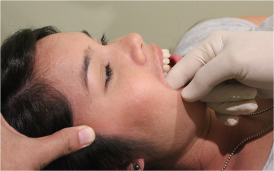
The muscles usually injected in TMD are the temporalis and masseter as shown in figure 1. There may be clinically relevant trigger points in other surrounding muscles such as the frontalis, occipitalis, trapezius, levator scapula, sternocleidomastoid, semispinalis capitis, splenius cervicis, and trapezius which may also be injected.[22]

- Figure 1. Trigger point injections in the temporalis and masseter as described by Robbins et al.[22] Arrows show pain referral patterns, and dashed lines show pain destinations. The needles show common injection sites. The muscles can be located with gentle mouth opening and closing. For injections into the Occipitalis, Frontalis and Temporalis, the temporal artery must be located and then one finger placed over it, with the needle angled upwards to avoid puncture. Injection over three sites in the muscle may be required. For injection into the masseter, the needle is directly towards the posterior aspect of the ramus of the mandible.
Multiple injectable agents have been described and include local anaesthetics, corticosteroids, botulinum toxin, and dry needling. There is no strong evidence to support any one agent .[22] Regardless, the use of local anaesthetic is the most commonly utilised in practice and in the literature. This is normally 1% lidocaine or 0.5% bupivacaine, however bupivacaine has a higher risk of myonecrosis.[22][24] Injections are typically repeated monthly over several sessions.[22]
The only true contraindication to local anaesthetic trigger point injections is allergy to the agent. Relative contraindications include vulnerability to vaso-vagal syncope, pregnancy (lignocaine is preferred), open skull defect, local or systemic infection, international normalised ratio (INR) over 3, and unclear anatomical landmarks.[22]
After locating the injection sites, the site is marked by applying pressure to the area with a retracted ballpoint pen. The areas are then cleaned with an alcohol swab or similar. The size of the needle depends on the depth of the muscle and is normally between 21 to 27g. A no-touch technique is utilised by pulling the surrounding skin taut with two fingers. The area is then injected between the two fingers, 1-1.5cm away from the injection site and advanced at 30-degree angle aiming towards the trigger point. The area is then aspirated, confirming the absence of a vessel at the needle point, and 0.1-0.3mL is injected per trigger point. The needle may be reangled through the same puncture site for injection of other nearby trigger points. A total volume of 2 to 4mLs can be used per session.[22]
Joint Injections
- Main article: TMJ Injection
There have been several promising older studies in the 80s and 90s looking at intra-articular steroid injections, and they appear to have a good safety profile.[25][26][27]
Injected high molecular weight hyaluronic acid has also been studied, and is used for visco-supplementation and its anti-inflammatory effects, with favourable results.[26][28][29][30][31]
One study compared directly the intra-articular injection of hyaluronic acid versus the corticosteroid betamethasone for TMJ osteoarthritis. There was no placebo or self-care group. There was improvement in pain and function in both groups at six months, with a greater response in the hyaluronic acid group.[32]
A typical protocol for these injections is 0.5mL of betamethasone or 0.5mL of hyaluronic acid injected into the upper joint space using the no-touch technique as described previously and then repeated one or two weeks later.[28][32] Hyaluronic acid infiltrations (arthrocentesis) repeated five times over a period of several weeks is another technique shown to be effective in disc displacement without reduction.[30]
Intraarticular dextrose prolotherapy (20%) repeated at 0, 1, and 2 months, has also been shown to have efficacy.[33]
Manual Therapy
Manual therapy is a physical treatment used to treat a variety of musculoskeletal complaints. It is subdivided into joint based (joint manipulation and mobilisation), soft tissue massage (deep tissue, trigger point among others), and neural based techniques. There is limited evidence for its effectiveness across a variety of conditions.[34] With regards to temporomandibular dysfunction, a systematic review in 2015 found that manual therapy was more effective than conservative treatment, resulting in improved mouth opening range of motion and decreased pain.[35]
Joint Mobilisation
Joint mobilisation is a core manual therapy technique used to treat a variety of musculoskeletal disorders by reducing pain and increasing range of motion. The technique utilised is determined based on the identified joint restriction. It should be used with caution in those with hypermobility.[36]
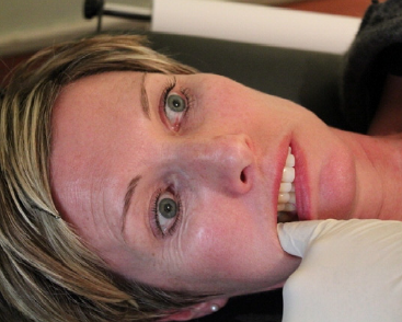
Taylor et al found in a small study that grade IV (small oscillatory movements at end range) mandibular mobilisation was effective for improving TMJ range of motion.[37] The practitioner’s thumb is placed over the ipsilateral lower molars, with the remaining fingers wrapped around the mandible. A grade IV caudal distraction mobilisation is then performed, by taking the joint into resistance and then applying oscillatory downward pressure through the thumb. This is done for 60 seconds for a total of three sets, with 10 seconds rest in between (Figure 2).

- Figure 2. Positioning for mandibular distraction techniques. Note that the practitioner stands on the contralateral side, with the contralateral thumb pad applied to the lower molars, and the ipsilateral hand is monitoring the TMJ. The patient’s head is resting on the practitioner’s chest. This can also be performed with the patient prone.[38]
A randomised controlled trial of those with TMD with disc displacement compared home mandibular distraction against splint therapy. There was improvement in both groups, but the exercise group had a statistically significant greater improvement in function. The exercise protocol was similar: there was an initial warmup with several small amplitude mouth opening and closing movements, followed by the patient pushing caudally on the mandibular teeth to the point of pain and then holding for 30 seconds for a total of 4 sets per day.[39]
The jaw can also be mobilised in other ranges such as anteriorly, medially, laterally, and caudal-anterior-medial (CAM) depending on the located restriction. The principles remain the same as per the inferiorly directed technique. For the anterior glide, the thumb pulls the jaw anteriorly while the contralateral hand provides a posterior stabilisation force. With medial, lateral, and CAM mobilisation, the directional force is applied with the palm of the hand over the ramus of the jaw, while the contralateral hand provides a stabilisation force through the contralateral zygomatic arch.[36]
Self-mobilisation with a medially directed force has been described by Shaffer et al to be a useful home exercise, and is summarised in figure 3.[36]

- Figure 3. Self-mobilisation of the TMJ from Shaffer et al.[36] In these home exercise examples, the large arrow signifies the application of a medially directed force through the mandibular ramus or condyle, while the contralateral hand applies a stabilisation force about the zygomatic arch. The technique is performed with both mouth-closed (left) and mouth-opened (right).
Mobilisation with Movement
Mobilisation with movement can also be performed to the TMJ. For example the clinician can perform an anteromedial glide of mandible on stabilised temporal bone with jaw opening. Ensure the glide pressure is applied close to the TMJ, not on the posterior aspect of the mandible.
Postural and other Therapeutic Exercises
Forward head posture is sometimes theorised to be a potential causative factor in TMD. The hypothetical basis is that forward head posture with rounded shoulders creates abnormal muscular balance and the translation of the mandibular postero-superiorly therefore causes increased pressure of the condyle onto the disc and glenoid fossa.[11][40]
The Rocabado protocol (Figure 4) is a home exercise program, used in conjunction with self-care. It has the aim of improving postural position thereby restoring normal muscle and joint relationships. The program includes 6 exercises which are performed 6 times each and 6 times a day.[11]
There have been conflicting results with regards to the efficacy of postural exercises. One randomised trial found it to be inferior to self-care, and showed that it did not in fact improve head posture.[11] However another study showed the opposite conclusion with the superiority of postural exercises over self-care.[41] More studies are likely needed in this area.

- Figure 4. The 6x6 Rocabado home exercise program.[11]
There is no evidence that weak muscles are related to TMD, nor is there evidence to dictate which exercises are best. It is important to note that most patients with TMD over-activate their masticatory muscles and so relaxation techniques may in fact be more useful.[36] This concept of the importance of muscle relaxation and the counterproductive effects of strengthening has been studied in other musculoskeletal disorders such as low back pain.[42]
Myofascial Therapy
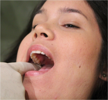
Intraoral myofascial therapy for myogenic TMD has been studied by Kalamir et al and shown to be more effective than wait list control and self-care.[43][44] The treatment approach is summarised in Figure 5. The strength of the patient’s gag reflex limits utility. Extra-oral friction massage can also be useful when applied to the affected fibres of the temporalis muscle.[36]
Intra-oral temporalis tendon release. Light pressure is applied to the temporalis tendon insertion on the coronoid, while the contralateral hand applies extra-oral longitudinal pressure along the anterior fibres of the temporalis muscle. The patient incrementally opens their mouth to maximum range.
Intra-oral medial and lateral pterygoid release. Posterior and cranial pressure is applied to the pterygoid muscles, maintained for 5 seconds.
Sphenopalatine ganglion technique. The fifth finger is inserted along the buccal surface while the patient lightly occludes their teeth, moving slowly posteriorly until the sphenopalatine fossa is palpated. The patient then lifts their head off the table pressing into the examiner’s finger. This is repeated 3 times, then the patient relaxes onto the bed and gentle pressure is applied to the masseter and pterygoids. Ipsilateral eye watering may occur.


Psychological Therapies
Cognitive behavioural therapy (CBT) is the most commonly used psychological approach for treating chronic pain, with its aim being to reduce maladaptive behaviour. Patients learn how their automatic thought processes contribute to their symptoms, how to challenge these thoughts, relaxation techniques, and discussion of fear-avoidance behaviour among other strategies.[45] A Cochrane review assessing CBT for the treatment of chronic pain concluded that CBT compared to treatment as usual had weak effects on improving pain and disability and was effective at improving psychological outcomes.[46]
Patients with TMD often have associated psychological symptoms and therefore CBT has been studied a treatment option. However, Liu et al in their meta-analysis found significant numerical inconsistencies in five of the trials they assessed, concluding that there is only weak evidence to support its implementation for the reduction in TMJ related pain and function.[47] Pragmatically, as a minimally-invasive therapeutic option, CBT could be potentially be considered as an adjuvant therapy for when there are significant co-existent psychological factors.
Occlusive Splints
Occlusion is the dental term for the bite. Many patients with TMD have poor occlusion, and it is theorised that this could be a contributing factor in some people to pain and disability due to the resulting imbalance in masticatory muscle activity. An occlusion splint, fitted by a dentist, is used at night and helps achieve an anatomically aligned disc-condyle position, thereby reducing this abnormal muscle activity (Figure 6).
Ebrahim et al in 2012 found low to moderate quality evidence for reduction in pain in their systematic review of 11 trials.[48] Since that publication, a randomised trial in 2015 found no improvement in pain compared to counselling and masticatory muscle exercises at one year follow up.[49] In addition an 8 year follow up trial published in 2013 found that more than 75% reported an improvement in TMJ pain, but there was no difference between the intervention and control groups.[50] Owing to their cost and inconsistent evidence, it is the author’s view that occlusive splints should not be considered first line.

- Figure 6. Schematic of how an occlusion splint improves alignment of the TMJ. Image from BiteFx.[51]
Conclusion
TMD is a common musculoskeletal complaint and can involve pain and dysfunction of the TMJ itself, the surrounding muscles, or both. Cervical spine dysfunction and psychosocial stressors are also often present.[6]
The first line treatment is usually self-care. There are no trials assessing this intervention against waitlist control, however it is low risk and many patients improve spontaneously. If this is insufficient other non-surgical strategies may be considered. The interventions with the best evidence base include joint mobilisation, myofascial therapy, and injections. Larger studies are required to confirm previous findings. Other interventions to be considered with a mixed evidence base include pharmacological therapy, home exercises, occlusive splints, and psychological therapies. Access to these interventions will depend on local expertise and cost.
Ultimately the clinician will be guided by their clinical assessment and may utilise multiple treatment modalities to improve pain and function.[36]
References
This article is based off Jeremy Steinberg's MSM709 university essay.
- ↑ Facial Pain | National Institute of Dental and Craniofacial Research [Internet]. [cited 2018 Nov 1]. Available from: https://www-nidcr-nih-gov/research/data-statistics/facial-pain
- ↑ 2.0 2.1 2.2 2.3 2.4 2.5 Goddard G, Mauro G. Temporomandibular disorders, a review of current diagnosis and treatment. Dent Cadmos. 2018 May;86(05):364.
- ↑ 3.0 3.1 3.2 Schiffman EL, Ohrbach R, Truelove EL, Feng T, Anderson GC, Pan W, et al. The Revised Research Diagnostic Criteria for Temporomandibular Disorders: Methods used to Establish and Validate Revised Axis I Diagnostic Algorithms. J Orofac Pain. 2010;24(1):63–78.
- ↑ Mehta NR, Forgione AG, Rosenbaum RS, Holmberg R. “TMJ” triad of dysfunctions: a biologic basis of diagnosis and treatment. J Mass Dent Soc. 1984;33(4):173–6, 212–3.
- ↑ Bayat M, Abbasi AJ, Noorbala AA, Mohebbi SZ, Moharrami M, Yekaninejad MS. Oral health-related quality of life in patients with temporomandibular disorders: A case-control study considering psychological aspects. Int J Dent Hyg. 2018 Feb;16(1):165–70.
- ↑ 6.0 6.1 6.2 6.3 Shaffer SM, Brismée J-M, Sizer PS, Courtney CA. Temporomandibular disorders. Part 1: anatomy and examination/diagnosis. J Man Manip Ther. 2014 Feb;22(1):2–12.
- ↑ Ren YF, Isberg A. Tinnitus in patients with temporomandibular joint internal derangement. Cranio J Craniomandib Pract. 1995 Apr;13(2):75–80.
- ↑ Schiffman E, Ohrbach R. Executive Summary of the Diagnostic Criteria for Temporomandibular Disorders (DC/TMD) for Clinical and Research Applications. J Am Dent Assoc 1939. 2016 Jun;147(6):438–45.
- ↑ Okeson JP, Perez C, Fricton JR. Temporomandibular Joint Disorders. In: Ferreira JNAR, Fricton J, Rhodus N, editors. Orofacial Disorders: Current Therapies in Orofacial Pain and Oral Medicine [Internet]. Cham: Springer International Publishing; 2017 [cited 2018 Nov 4]. p. 145–57. Available from: https://doi.org/10.1007/978-3-319-51508-3_14
- ↑ 10.0 10.1 10.2 Story WP, Durham J, Al-Baghdadi M, Steele J, Araujo-Soares V. Self-management in temporomandibular disorders: a systematic review of behavioural components. J Oral Rehabil. 2016 Oct;43(10):759–70.
- ↑ 11.0 11.1 11.2 11.3 11.4 Mulet M, Decker KL, Look JO, Lenton PA, Schiffman EL. A randomized clinical trial assessing the efficacy of adding 6 x 6 exercises to self-care for the treatment of masticatory myofascial pain. J Orofac Pain. 2007;21(4):318–28.
- ↑ 12.0 12.1 Michelotti A, Steenks MH, Farella M, Parisini F, Cimino R, Martina R. The additional value of a home physical therapy regimen versus patient education only for the treatment of myofascial pain of the jaw muscles: short-term results of a randomized clinical trial. J Orofac Pain. 2004;18(2):114–25.
- ↑ Mujakperuo HR, Watson M, Morrison R, Macfarlane TV. Pharmacological interventions for pain in patients with temporomandibular disorders. Cochrane Database Syst Rev. 2010 Oct 6;(10):CD004715.
- ↑ Ekberg EC, Kopp S, Akerman S. Diclofenac sodium as an alternative treatment of temporomandibular joint pain. Acta Odontol Scand. 1996 Jun;54(3):154–9.
- ↑ Ta LE, Dionne RA. Treatment of painful temporomandibular joints with a cyclooxygenase-2 inhibitor: a randomized placebo-controlled comparison of celecoxib to naproxen. Pain. 2004 Sep 1;111(1):13–21.
- ↑ Herman CR, Schiffman EL, Look JO, Rindal DB. The effectiveness of adding pharmacologic treatment with clonazepam or cyclobenzaprine to patient education and self-care for the treatment of jaw pain upon awakening: a randomized clinical trial. J Orofac Pain. 2002;16(1):64–70.
- ↑ Cascos-Romero J, Vázquez-Delgado E, Vázquez-Rodríguez E, Gay-Escoda C. The use of tricyclic antidepressants in the treatment of temporomandibular joint disorders: systematic review of the literature of the last 20 years. Med Oral Patol Oral Cirugia Bucal. 2009 Jan 1;14(1):E3-7.
- ↑ Tchivileva et al.. Efficacy and safety of propranolol for treatment of temporomandibular disorder pain: a randomized, placebo-controlled clinical trial. Pain 2020. 161:1755-1767. PMID: 32701836. DOI. Full Text.
- ↑ Lobbezoo F, van Denderen RJ, Verheij JG, Naeije M. Reports of SSRI-associated bruxism in the family physician’s office. J Orofac Pain. 2001;15(4):340–6.
- ↑ Winocur E, Gavish A, Halachmi M, Eli I, Gazit E. Topical application of capsaicin for the treatment of localized pain in the temporomandibular joint area. J Orofac Pain. 2000;14(1):31–6.
- ↑ Alvarez DJ, Rockwell PG. Trigger points: diagnosis and management. Am Fam Physician. 2002 Feb 15;65(4):653–60.
- ↑ 22.0 22.1 22.2 22.3 22.4 22.5 22.6 22.7 22.8 Robbins MS, Kuruvilla D, Blumenfeld A, Charleston L, Sorrell M, Robertson CE, et al. Trigger Point Injections for Headache Disorders: Expert Consensus Methodology and Narrative Review. Headache J Head Face Pain. 2014 Oct 1;54(9):1441–59.
- ↑ Karadaş Ö, Gül HL, Inan LE. Lidocaine injection of pericranial myofascial trigger points in the treatment of frequent episodic tension-type headache. J Headache Pain. 2013 May 22;14:44.
- ↑ Blumenfeld A, Ashkenazi A, Grosberg B, Napchan U, Narouze S, Nett B, et al. Patterns of use of peripheral nerve blocks and trigger point injections among headache practitioners in the USA: Results of the American Headache Society Interventional Procedure Survey (AHS-IPS). Headache. 2010 Jun;50(6):937–42.
- ↑ Wenneberg B, Kopp S, Gröndahl HG. Long-term effect of intra-articular injections of a glucocorticosteroid into the TMJ: a clinical and radiographic 8-year follow-up. J Craniomandib Disord Facial Oral Pain. 1991;5(1):11–8.
- ↑ 26.0 26.1 Kopp S, Carlsson GE, Haraldson T, Wenneberg B. Long-term effect of intra-articular injections of sodium hyaluronate and corticosteroid on temporomandibular joint arthritis. J Oral Maxillofac Surg Off J Am Assoc Oral Maxillofac Surg. 1987 Nov;45(11):929–35.
- ↑ Kopp S, Wenneberg B. Effects of occlusal treatment and intraarticular injections on temporomandibular joint pain and dysfunction. Acta Odontol Scand. 1981;39(2):87–96.
- ↑ 28.0 28.1 Hepguler S, Akkoc YS, Pehlivan M, Ozturk C, Celebi G, Saracoglu A, et al. The efficacy of intra-articular sodium hyaluronate in patients with reducing displaced disc of the temporomandibular joint. J Oral Rehabil. 2002 Jan;29(1):80–6.
- ↑ Guarda-Nardini L, Tito R, Staffieri A, Beltrame A. Treatment of patients with arthrosis of the temporomandibular joint by infiltration of sodium hyaluronate: a preliminary study. Eur Arch Oto-Rhino-Laryngol Off J Eur Fed Oto-Rhino-Laryngol Soc EUFOS Affil Ger Soc Oto-Rhino-Laryngol - Head Neck Surg. 2002 May;259(5):279–84.
- ↑ 30.0 30.1 Guarda-Nardini L, Masiero S, Marioni G. Conservative treatment of temporomandibular joint osteoarthrosis: intra-articular injection of sodium hyaluronate. J Oral Rehabil. 2005 Oct;32(10):729–34.
- ↑ Yeung RWK, Chow RLK, Samman N, Chiu K. Short-term therapeutic outcome of intra-articular high molecular weight hyaluronic acid injection for nonreducing disc displacement of the temporomandibular joint. Oral Surg Oral Med Oral Pathol Oral Radiol Endod. 2006 Oct;102(4):453–61.
- ↑ 32.0 32.1 Bjørnland T, Gjaerum AA, Møystad A. Osteoarthritis of the temporomandibular joint: an evaluation of the effects and complications of corticosteroid injection compared with injection with sodium hyaluronate. J Oral Rehabil. 2007 Aug;34(8):583–9.
- ↑ Louw et al.. Treatment of Temporomandibular Dysfunction With Hypertonic Dextrose Injection (Prolotherapy): A Randomized Controlled Trial With Long-term Partial Crossover. Mayo Clinic proceedings 2019. 94:820-832. PMID: 30878157. DOI.
- ↑ Clar C, Tsertsvadze A, Court R, Hundt GL, Clarke A, Sutcliffe P. Clinical effectiveness of manual therapy for the management of musculoskeletal and non-musculoskeletal conditions: systematic review and update of UK evidence report. Chiropr Man Ther. 2014 Mar 28;22(1):12.
- ↑ Martins WR, Blasczyk JC, Aparecida Furlan de Oliveira M, Lagôa Gonçalves KF, Bonini-Rocha AC, Dugailly P-M, et al. Efficacy of musculoskeletal manual approach in the treatment of temporomandibular joint disorder: A systematic review with meta-analysis. Man Ther. 2016 Feb 1;21:10–7.
- ↑ 36.0 36.1 36.2 36.3 36.4 36.5 36.6 Shaffer SM, Brismée J-M, Sizer PS, Courtney CA. Temporomandibular disorders. Part 2: conservative management. J Man Manip Ther. 2014 Feb;22(1):13–23.
- ↑ Taylor M, Suvinen T, Reade P. The effect of Grade IV distraction mobilisation on patients with temporomandibular pain-dysfunction disorder. Physiother Theory Pract. 1994 Jan 1;10(3):129–36.
- ↑ TMJ caudal anterior glide - YouTube [Internet]. [cited 2018 Nov 4]. Available from: https://www.youtube.com/watch?v=IF_PDLU78dY
- ↑ Haketa T, Kino K, Sugisaki M, Takaoka M, Ohta T. Randomized clinical trial of treatment for TMJ disc displacement. J Dent Res. 2010 Nov;89(11):1259–63.
- ↑ Gonzalez HE, Manns A. Forward head posture: its structural and functional influence on the stomatognathic system, a conceptual study. Cranio J Craniomandib Pract. 1996 Jan;14(1):71–80.
- ↑ Wright EF, Domenech MA, Fischer JR. Usefulness of posture training for patients with temporomandibular disorders. J Am Dent Assoc 1939. 2000 Feb;131(2):202–10.
- ↑ Lederman E. The myth of core stability. J Bodyw Mov Ther. 2010 Jan 1;14(1):84–98.
- ↑ 43.0 43.1 Kalamir A, Bonello R, Graham P, Vitiello AL, Pollard H. Intraoral myofascial therapy for chronic myogenous temporomandibular disorder: a randomized controlled trial. J Manipulative Physiol Ther. 2012 Jan;35(1):26–37.
- ↑ 44.0 44.1 Kalamir A, Graham PL, Vitiello AL, Bonello R, Pollard H. Intra-oral myofascial therapy versus education and self-care in the treatment of chronic, myogenous temporomandibular disorder: a randomised, clinical trial. Chiropr Man Ther. 2013 Jun 5;21(1):17.
- ↑ Ehde DM, Dillworth TM, Turner JA. Cognitive-behavioral therapy for individuals with chronic pain: Efficacy, innovations, and directions for research. Am Psychol. 2014;69(2):153–66.
- ↑ Williams AC de C, Eccleston C, Morley S. Psychological therapies for the management of chronic pain (excluding headache) in adults. Cochrane Database Syst Rev [Internet]. 2012 [cited 2018 Sep 22];(11). Available from: https://www.cochranelibrary.com/cdsr/doi/10.1002/14651858.CD007407.pub3/abstract
- ↑ Liu HX, Liang QJ, Xiao P, Jiao HX, Gao Y, Ahmetjiang A. The effectiveness of cognitive-behavioural therapy for temporomandibular disorders: a systematic review. J Oral Rehabil. 2012 Jan;39(1):55–62.
- ↑ Ebrahim S, Montoya L, Busse JW, Carrasco-Labra A, Guyatt GH, Medically Unexplained Syndromes Research Group. The effectiveness of splint therapy in patients with temporomandibular disorders: a systematic review and meta-analysis. J Am Dent Assoc 1939. 2012 Aug;143(8):847–57.
- ↑ Qvintus V, Suominen AL, Huttunen J, Raustia A, Ylöstalo P, Sipilä K. Efficacy of stabilisation splint treatment on facial pain - 1-year follow-up. J Oral Rehabil. 2015 Jun;42(6):439–46.
- ↑ Erixon CL, Ekberg E. Self-perceived effects of occlusal appliance therapy on TMD patients: an eight-year follow-up. Swed Dent J. 2013;37(1):13–22.
- ↑ Dental occlusion patient education software-used by dental educators [Internet]. [cited 2018 Sep 23]. Available from: https://www.bitefx.com/
Literature Review
 Literature Review
- Reviews from the last 7 years: review articles, free review articles, systematic reviews, meta-analyses, NCBI Bookshelf
- Articles from all years: PubMed search, Google Scholar search.
- TRIP Database: clinical publications about evidence-based medicine.
- Other Wikis: Radiopaedia, Wikipedia Search, Wikipedia I Feel Lucky, Orthobullets,

