Tendinopathy
Tendinopathy is the term for persistent tendon pain and loss of function related to mechanical loading. Tendon tears are macroscopic discontinuity of a load-bearing tendon that can be partial or complete.[1] Also see article on Calcific Tendinopathy, which is a different disease process.
Pathology

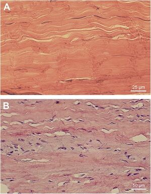
Tendinopathy is a tendon disorder of tissue remodelling that occurs in a continuum across the major tendons of the body. It is characterised by certain changes within the tendon leading to a reduction in tensile load bearing potential. It can can lead to pain and impaired function. Tendon injury can occur in the mid-portion of the tendon (e.g. mid-portion achilles tendon) or more commonly at the enthesis insertion site to bone (e.g. insertional-portion achilles tendon, patellar tendon, and elbow tendons). The key treatment principal is progressive careful load management.
There are profound changes in tendon structure in tendinopathy. In tendinopathy there is loss in capacity to take load through an area of degeneration. This leads to difficulty in stimulating any reparative response.
It isn't possible to tear normal collagen. With tendon rupture there is rupture of highly degenerative tendon without much normal tissue. Therefore without a collagen tearing response, there is no bleeding response, and without bleeding there is no inflammatory response.
The tendon cells, called tenocytes, are intimately connected to collagen and are sensitive and responsive to matrix load. The mechano-set point of the cell is dependent on the load that is placed on it, and this can be up- or down-regulated, with positive and negative responses.
Inflammation
inflammation occurs in tendinopathy, however it is low grade. There are a few inflammatory cells peri-vascularly, and also some inflammatory mediators that are possibly produced by tenocytes and visiting cells.
Mast cells are recruited to the tendon tissue possibly due to substance P release from tenocytes or peripheral nerve endings. Substance P is known to be involved in chemical nociception and neurogenic inflammation. Mast cells can degranulate in response to substance P releasing histamine, tryptase and other substances. Tryptases and other proteases that are released activate protease-activated receptors (PARs) which are involved in fibrosis, hyperalgesia and neovascularisation.[2]
It is thought that inflammation is unlikely to be the prime driver of pathology or pain, and in the past when management was directed at this, outcomes were poor. While with rupture or bleeding in a tendon, inflammation is a prime driver.
Terminal state
Degenerative tendinopathy is a terminal state and cannot be recovered. In early stages of tendon pathology it is reversible, however once the tissue is in a non-load bearing state, there is no capacity to repair. Tendon healing and repair requires load sensitivity and a normal cell. In a degenerative tendon there is no collagen structure, and the pathology is therefore mechanically "deaf" and receives no stimulus to repair. The cells are also chondrocytic, they undergo metaplasia from a tenocyte to a chondrocyte, and produce a lot more cartilage based proteoglycans and type III as well as type II collagen. Therefore the normal structure cannot be recovered. However tendon pathology is not passive, it has a metabolic rate 25 times higher than a normal tendon, but it has no stimulus to create structure, and so this is all in vain.
Reactive change
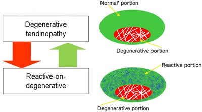
A tendon with degeneration will also have an area of normal tendon. So with rehabilitation when there is overload placed only on the normal tendon, a reactive change occurs in the normal tendon. This is called reactive on degenerative tendinopathy. The normal tendon around a degenerative area protects the tendon by structuring more normal tissue. Most individuals with degeneration form enough normal tendon to allow the structure to take load, in fact usually even more than a non-degenerative tendon. This relates to the management principle of "treat the doughnut not the hole." It also counters the theory that regenerative procedures such as platelet rich plasma injections are needed, as the body has already done this.
Tendon pain
Tendon pain only occurs in the presence of pathology. Pathology increases the risk of pain 3-7 times, however pathology can occur without pain. The nociceptive driver is unknown, but it may be the cell, and not due to inflammation or neovascularisation. The sensory innervation of the tendon is quite peripheral, in the peritendon, muscle, and bone-tendon junctions. There is little to no innervation within the tendon due to the massive loads placed on the tendon, which would theoretically cause pain even with a normal structure.
There are motor and sensory changes consistent with nociception, with changes in excitability and inhabitation to the affected muscle of the painful tendon. The changes all relate to motor-drive, and there is no evidence of any central or nociplastic drive to pain. Tendinopathy is a nociceptive condition.
Pain, poor function, and pathology/poor structure are three different concepts that often overlap, but can also exist on their own.
Continuum Model


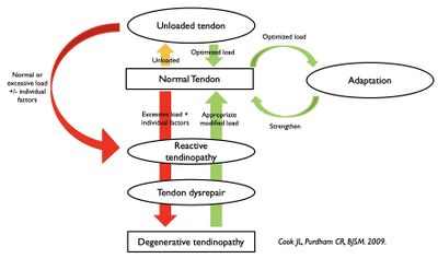
We can now talk about the continuum model.[3] There appears to be a continuum of tendon pathology explaining the clinical presentations of load induced tendinopathies. There have been three proposed stages: reactive tendinopathy, tendon dysrepair, and degenerative tendinopathy
Reactive Tendinopathy
Reactive tendinopathy is seen in acute overload (e.g. dramatic increase in activity levels) and after a direct blow (e.g. a fall onto the patellar tendon). The most vulnerable tendons are the Achilles, patellar, and elbow.
There is a non-inflammatory proliferative response in the cell and matrix following an acute tensile or compressive overload. This leads to a short term adaptive response involving tendon thickening to reduce stress through an increase in the cross-sectional area. This process stands in contrast to normal tendon adaptation to tensile load where we see stiffening without much change in thickness.
At the cellular level there is metaplasia of the tenocytes to become more chondroid in shape with more production of proteoglycans and subsequent increase in water content. The collagen integrity is largely maintained, but there can be some longitudinal separation. The effect is a rapid increase in tendon thickness but with the capacity to revert to normal if overload is addressed.
On imaging there is a swollen fusiform tendon. On ultrasound there are intact collagen fibres with diffuse hypoechogenicity between the collagen due to the increase in water bound proteoglycans. There may not be many changes on MRI at this stage.
Tendon Dysrepair
This is generally seen in chronically overloaded tendons.
Here there is attempted tendon healing that is characterised by an increase in chondrocytes and myofibroblasts. There is an associated marked increase in the production of proteoglycans and collagen. The proteoglycan content leads to separation of the collagen and disorganisation of the matrix. The changes tend to be more focal with more matrix changes than the reactive stage. There may be neovascularisation and neuronal ingrowth. There is still some possibility of reversibility with careful load management.
On imaging the tendons are swollen and there is signs of disorganisation of the collagen with discontinuity and focal areas of hypoechogenicity. On doppler there may be vascularity. MRI shows increased signal in a swollen tendon.
Degenerative Tendinopathy
This is often seen in the middle-aged and older population but can be seen in younger individuals with chronically overloaded tendons. The Achilles tendon is the classic site in the middle-aged individual.
There is cell death (from apoptosis, trauma, tenocyte exhaustion) with a resultant increase in acellularity. We see a disorganised matrix with little collagen content and the in-filling with vessels. There may be islands of degenerative tendon between tendon at other stages as well as normal tendon. There is little capacity for reversible change, and rupture can occur if the changes are extensive enough or if placed under high enough load.
On imaging there are hypoechoic regions with little reflection from collagen. On doppler there are numerous and large vessels. On MRI there is increased signal and tendon size. The changes tend to be focal.
Biomechanics
Key tendon functions
- Energy storage and release. Tendons are viscoelastic. With slow stretching the tendon gets longer and doesn't store energy. In order to store energy on the tendon the tensile loading needs to be fast.
- Compression. Compression occurs just proximal to the tendon insertion. For example with the achilles tendon, there is compression of the tendon against the back of the calcaneum with dorsiflexion. Any hagland 'deformity' actually reduces the load on this tendon.
- Friction. This where the tendon moves too much through the peritendon structures, leading to a peritendon response.
There is only one true tendinopathy in the lower limb. The rest are enthesopathies and are prone to compressive loads, the best representation of that is gluteus medius/minimus tendon pain.
Clinical Features
Tendon pain is local, aggravated by load, and eased by unloading. Tendon pain can completely destroy the kinetic chain, and so in that way it can be very different to pain in other structures.
Pain is directly linked to overload. Overload can be a one off event, leading to an acute tendon tendon response - either reactive or reactive on degenerative. It will usually settle quickly if its only a single load.
With ongoing tendon pain, there is often a drive to stop loading the tendon, leading to negative consequences with loss of power, strength, and endurance. The reduced capacity means high load on the tendon with less activity, and therefore more pain.
Part of the clinical assessment is around working out which loads are clinically relevant - energy storage and release, compression, and/or friction.
Diagnosis is based on imaging abnormality and palpation tenderness, however neither of these are diagnostic of tendon pain.
In the setting of tendon rupture, two thirds never had pain.
Differentiating Lower Limb Tendinopathy
Tendon Profiles
Most tendons have a clear clinical profile such as the patellar and gluteus medius tendons, while the achilles tendon is harder to fit into a neat category. Athletes present with true tendinopathies, as tendinopathy is a condition of power athletes. Up to 40% of athletes have pathology but only a small number have symptoms. To some extent tendinopathy is a condition of older age but this is due to load exposure throughout life rather than age itself, and so highly active young athletes can have tendinopathy.
Patellar tendinopathy - young men, jumping sport, occasionally in elite female athletes but half the rate as men. It is common in adolescence if the tendon is loaded excessively before the end of puberty, similar to Osgood Schlatter in the distal patellar tendon. This pathology is carried with the individual through life with a permanent central cyclops lesion which may or may not give tendon pain. This is over-diagnosed as a cause of anterior knee pain, and the correct diagnosis is usually patellofemoral pain syndrome. Patellofemoral pain can generate pain over the tendon, and so pain inferior to the patellofemoral joint can still be patellofemoral pain. Fat pad irritation as a diagnosis is controversial.
| Patellar tendinopathy | Patellofemoral pain |
|---|---|
| Localised pain that doesn't move | Pain can be in the tendon but is less specific |
| Specific tasks aggravate pain (only jumping etc) | Vaguer tasks aggravate pain (low load tasks, pain when not loading) |
| Muscle wasting, especially quads, and also calf | Less muscle change |
| Stiff hop which is of poor quality and patient won't bend the knee | Soft hop due to poor hip control |
Gluteal tendinopathy - post-menopausal women, very occasionally in men. There is compression in women not men due to the broader pelvis in women. It is rare to find an intact tendon in women over the age of 60. Bursitis can't be a diagnosis in isolation. Bursal involvement means that there is an enthesopathy. Overall diagnosing this condition is often difficult due to overlapping pain regions. Reduced pain with isometric loading (single leg hip hitch) can be helpful in making the diagnosis in the clinic.
Adductor tendinopathy - This is difficult to diagnose due to multiple sources of pain. There is no cortical bone between bone and tendon. More common in men.
Achilles tendinopathy - non-homogenous presentation. There is loading across life, with simple everyday tasks being energy storage tasks such as walking up and down stairs. Therefore relatively sedentary people can present with this. There may also be an endurance capacity issue. Mid-portion tendinopathy can be confused with plantaris tendinopathy, posterior joint problems, peritendon problems. The superficial bursa can be involved in isolation and can be confused with insertional tendinopathy. Sural nerve irritation is another diagnostic possibility.
Plantaris tendinopathy - often chronic non-responsive achilles pain. Can be acute onset. Often the problem is a bit higher than typical for mid-portion achilles tendinopathy, and can be medial in acute stages. Patients don't like being in positions of dorsiflexion such as with flat shoes and bare feet as there is higher compression with more dorsiflexion. There may be a history of multiple "mild calf tears."
Quadriceps tendinopathy - this can be confused with superomedial knee plica.
Peritendon diagnoses - This relates to friction from sliding. There are limited clinical tests. There is inconsistent load dependent increase in pain. Patients are often have less pain with hoping. There may or may not be crepitus, and using a stethoscope can be helpful in hearing for this. Response to heparinoid cream can be used to make the diagnosis.
Retinacular conditions - This isn't usually seen in the lower limb and is due to impingement on the tendon. The classic condition in the upper limb is de Quervain's. In this condition the primary problem is the retinaculum, not the tendon. The retinacular changes occur first with compression on the peritendon. Long term peritendon changes can subsequently induce tendon pathology. In the lower limb it can be seen in the Achilles with the posterior retinaculum.
Systemic disease - Enthesopathies are seen in spondyloarthropathies. Insulin resistance and type 2 diabetes are risk factors for tendon problems. Pes anserinus problems should spur looking for systemic disease. Problems are often bilateral.
There can be a combination of pain sources, with tendon pain being only part of the problem. Overall it does tend to be mostly the tendon or not at all. In the patellar tendon we don't see a mix of nociceptive input.
Tenderness
Tenderness is a poor diagnostic sign. Tenderness relates to how much the tendon has been used, and isn't linked to either symptoms or imaging findings. Tenderness is likely to persist even with recovery in pain and function, and so don't allow the patient to poke it as it isn't an indicator of progress. The patellar tendon is the most painful area to palpation in the knee in both osteoarthritis and patellofemoral joint pain. This doesn't correlate with pain provocation testing (decline squat), or imaging.
Cardinal signs
- The pain is related to some sort of overload. This can be quite obvious with a dramatic change in load or very subtle such as a change in shoes.
- The pain is localised, one or maximum two fingers in area. If it is a broad are of pain it either isn't tendon pain or there is an additional area of nociception.
- There is a load dependent increase in pain. The pain stays localised with increasing load. With an increase in the load on the tendon and the rate of loading there is an increase in pain. For example in achilles tendinopathy, a double leg calf raise may give no pain, a single leg calf raise may give a small amount of pain, a double leg jump may give a greater pain, and a single leg hop will be the most painful.
- The pain is worse with energy storage and release loads. There may be an inability to store energy in the tendon, for example inability to hop.
- The pain is worse the day after high loads. There is an inability to recover from load for >24 hours, or up to 48 hours in severe cases.
- Specific tendon signs - such as sitting pain with hamstring, side lying pain with gluteus medius, etc.
Cardinal overloads
- Energy storage and release loads. Such as jumping, sprinting, changing direction.. Achilles and gluteal tendons can experience these loads with everyday activities.
- Compression. The load at length such at sprint take offs with ankle dorsiflexion.
- Friction. Excessive and large movements. Seen in the long tendons of the hand and foot.
Staging
Reactive tendinopathy is usually seen in younger individuals (15-25 years). There is a rapid onset of pain that is generally related to load. The load substantially exceeds the tendon's previous exposure. There is a fusiform swelling of the tendon at 3-4cm from the insertion. The pain is easily aggravated by exercise, and is slow to settle. In adults reactive tendinopathy is uncommon and occurs in compressive loads such as a direct blow to the back of the achilles or elbow.
Reactive on degenerative tendinopathy is usually seen in older individuals (40 years plus). There is a past history with load related exacerbations. The onset occurs after overload. There is variable swelling, and the tendon is less irritable. This much more common than reactive tendinopathy.
Degenerative tendinopathy is seen in older individuals (30-60 years) with a long history of minimal symptoms. There is a variable history of swelling. The patient often uses unloading strategies and there may be atrophy. This category is not painful, is quite common, but is often not seen clinically due to the minimal pain.
Response to isometric loading
Here we look for an immediate response to isometric exercise confirming that the tendon is the source of the pain. Do 4 sets of up to 45 second holds, with 1-2 minute rest in between. This should at least halve the pain. If this provokes the pain then non-tendinopathic causes of pain are more likely. For example in anterior knee pain, do isometric leg extensions, and if this provokes the pain then the patients problem is likely patellofemoral pain.
Taping
Using diamond taping over the the anterior knee can help differentiate between patellofemoral pain and patellar pain. Taping should reduce patellofemoral load pain, while the pain stays the same if the problem is the tendon
Complicating factors
Previous injections can complicate matters. Prolonged unloading affects all function and so this can be difficult to tease out. Other than these two factors there are no other real complicating factors. Specifically, there is no evidence of "central sensitisation" in the lower limb. Tendinopathy is a nociceptive condition not a nociplastic one.
Imaging
Imaging cannot diagnose the source of the problem, and also does not assist in predicting the prognosis. Imaging can also lead to harms as terms like tear and degeneration can result in kinesiophobia.
Much of what is seen on imaging is subjective with poor interobserver reliability. This includes amount of disorganisation, doppler flow, and intratendinous tears. In ultrasound an MR imaging of gluteal tendon pathology, using surgical/histological findings as the gold standard, there is good-to-excellent reliability for ultrasound in diagnosing full-thickness tears. However there is extremely poor reliability for tendinosis and partial tears for both MRI and US.[5]
A normal scan is highly predictive of the pain not coming from that tendon. However half of 'normal' tendons with pain develop pathology on imaging over one year. Abnormal imaging only means that the pain might be coming from the tendon, not that it is coming from there. There is a 3-18 times increase odds of having pain with an abnormal tendon on imaging.
Vascularity is not a cause of pain, but simply a marker of tendon degeneration.
We cannot identify the tendons that are at risk of rupture or poor outcomes either from imaging or clinical presentation.
The language used is very important. Explain to patients that they should not worry about the tear. This is because of the poor inter-observer reliability and that it rarely affects treatment. Also explain that a thick tendon is a good tendon, and shows that the tendon has compensated, and there is enough "doughnut" around the hole. The tendon isn't limiting them becoming pain-free but rather dysfunction is the problem.
Management

Management Approach Overview
- Ensure the tendon is the source of the pain. Imaging changes do not mean that the tendon is causing the pain. The wrong diagnosis will lead to the wrong treatment.
- Assess the existing muscle and tendon capacity and function
- Establish what the patient wants to achieve and the capacity and function required (e.g. sprinting athlete doesn't need endurance)
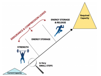
- Slowly build from one thing to the next, in very small steps. Improvement takes a long time and can't be rushed. Progress when there is stability in symptoms, and no flare with load.
- Initially unload the tendon from the tendon load that started to problem in order to control the pain and to get the correct loading on it.
- Use isometric exercises to reduce pain
- Address co-morbidities
- Use adjuncts as required.
Stage 1: Isometrics
Stage 2: Isotonics/heavy slow resistance
Stage 3: Energy Storage
Stage 4: Energy storage and release
Someone who understands tendons must set the program. Errors include incorrect or too little progressions, insufficient load (the exercise must be set at maximal tolerable load. If the load is too light then it is inefficient, if it is too heavy then you get increasing symptoms), too little attention to the kinetic chain, and following recipe programmes without addressing the individual.
An example is gluteal tendinopathy in a middle aged woman. Their current capacity is poor strength is all leg muscles, muscle wasting, poor endurance, and pain with simple loads. The required capacity is to be able to walk, shop, garden, and endurance. These patients won't be taken to energy storage and release loads. Rehabilitation will be focused on strength, usually with home-exercises, sit to stand, calf raises on two legs, isometric exercises etc. Commonly gluteal tendinopathy patients will be prescribed clams, but the load with clams is much too low and have virtually no effect.
Active Therapies
General
The general principles are exercise and correct loading. The goal of therapy is restoration of full pain-free function. In athletes there is the additional requirement of having the tendon be able to tolerate the highest load with energy storage and release. We also want the tendon to be able to tolerate loads other than tensile, mainly compression, and sometimes friction. The answer to improving pain is not changing structure.
Active therapy modalities are less likely to help when done in isolation.
Improvements in pain and function due to exercise are not linked to structural changes. Effective exercise acts on the normal part of the tendon, increasing its tolerance to load. Exercise also acts on the muscles, the kinetic chain, and the brain.
In an athlete, rehabilitation is not only about getting the tendon strong, but also getting it to a state where it can take on a high rate of tensile loading. Athletes with poor function will always have pain, and athletes with good function are protected from pain. There treatment is aimed at improving function, in order to lead to improvement in pain.
A big problem in the research in this area is that surrogate outcomes are usually used - a decrease in pain rather than looking at return to activity.
Active therapies often fail in the real world because it isn't perceived at cutting edge, it is slow, and it isn't expensive.
Isometric exercise
Isometric exercise acts on cortical inhibition, and there is relief for several hours if done correctly. It is more effective in the big muscle groups such as achilles, hamstrings, patella. Athletes are 20% stronger after isometrics. Isometric exercise also conditions the tendon.
It needs to be done with a long hold. 45 seconds for 5 repetitions, 1-2 times a day, with 2 minute rest between.
Eccentric exercise
Eccentric exercise loads the series elastic component of the tendon and muscle, i.e the spring. It doesn't strengthen the muscle, kinetic chain, change the brain, or address the individual. It doesn't adapt the tendon to energy storage loads. And it doesn't change compressive loads. The evidence is mixed and can be ineffective on their own, especially in athletes. Overall these should not be done in isolation, and not follow a set program.
Heavy slow resistance
This improves muscle strength, and the slow loads improve the mechanical strength of the tendon. Muscle endurance can be addressed with endurance sets. It doesn't fully address the kinetic chain and brain, and doesn't adapt the tendon to energy storage and release loads.
The weight needs to be heavy, but heavy for that specific person. It doesn't have to be using a squat rack. This is best done single leg, with each log. Once strength is obtained then can move on to adding function strength/endurance and energy storage and release.
Energy storage and release loading
Here you are adding speed by using plyometric exercises. This improves the loading rate capacity of the tendon. These are done 2-3 times a week only. Progress to sport based activities.
Load modification
Reduce the high loads placed on the painful tendon. For example sprinting and direction changing in an athlete. Tendon pain reduces athletic function as they can't jump, change direction, etc.
Reducing compression
In certain conditions the compressive load on the tendon should be reduced, for example with gluteus medius/minimus tendon pain.
Visual/auditory cues
Cues can improve cortical plasticity. An example is using a metronome.
Passive Therapies
Passive therapies should be an adjunct, and have to sit within the context of the load capacity and load tolerance of the tendon, muscle, kinetic chain, and brain. Passive therapy alone cannot result in improvement without an active intervention as well.
Injection therapies
Most injection therapies have a neural effect. Sclerosing injections are neurotoxic, and may be causing pain relief due to possible cytotoxic effects on the nociceptors. It likely doesn't matter what is injected, even saline has an effect, and so the effect of any injection is likely not acting on the pathology, but rather the neural drive.
PRP injection results in increased cell and GAG content, however this forces the tendon further away from normal, and there is no increase in collagen count. The tendon is already hypercellular and producing a large amount of GAGs. Needle tracts can be seen even months later on UTC scan, and so even the physical act of needle insertion into a tendon negatively affects tendon structure.
Topical nitroglycerin
Topical nitroglycerin has some evidence of utility. The exact mechanisms are unclear but it is thought that it may enhance collagen synthesis via the nitric oxide signalling molecule. It can be useful as an adjunct to physical therapy, and shouldn't be used on its own.
Nitroglycerin patches are placed directly over affected tendons. Generally one-fourth or one-half of a 5mg/24 hour or 10mg/24 hour patch is left on continuously for 24 hours. Patients should be advised that it can take 12 to 24 weeks to experience significant improvement. The skin should be monitored for signs of irritation and be made aware that dizziness and headache are potential side effects. Contraindications include hypotension, migraine headaches, rosacea, head injury, severe anaemia, and ingestion of phosphodiesterase inhibitors or oral or sublingual nitroglycerin.
For rotator cuff tendinopathy a systematic review that included four randomised trials out of 10 randomised trials in total (two of which weren't placebo controlled) found moderate to strong evidence that topical nitroglycerin reduces pain and improves mobility, strength, and function, compared to placebo at short term and 24 week follow up.[7]
For non-insertional achilles tendinopathy a randomised controlled trial found that topical nitroglycerin significantly reduced pain and improved function at 12 and 24 weeks. At six months, 78% of the intervention group tends, and 49% of the placebo group were asymptomatic, meaning it had an NNT of ~3.5 for this outcome. However, the mean effect size was 0.14 which is small.[8]
For patellar tendinopathy a controlled trial found no differences between groups over a 24 week period.[9] Likewise another trial of lateral elbow tendinopathy found no evidence with short term use but it only went to 8 weeks and didn't include resistance exercise.[10]
Topical heparinoid cream
Heparinoid cream (hirudoid) can be used for peritendon pain, however it is of uncertain benefit.
Shockwave therapy
Pain relief may be due to neuropraxic effects on the nerves. However shockwave therapy can actually negatively affect the structure for up to 6 weeks.
Rest
Rest can improve pain in the short term, but the long term consequences are profound. Rest is catabolic to the tendon. The tendon tissue loses structure and becomes pathological, and the mechanical strength of the tendon decreases in two weeks. There are also negative changes in the anti-gravity kinetic chain function and cortical motor drive. Subsequently after resting when initiating load again the pain is worse due to the rest. The strength of the tissue is only as great as the load placed on it. By decreasing load, you are decreasing tissue capacity.
Passive therapies such as injections come with prescribed rest, and so at least part of the beneficial effect on pain will be due to reducing load.
Surgery
Extra-tendinous surgery denervates the tendon, leading to pain relief, but not improving the load tolerance.
Intra-tendinous surgery results in an inflammatory proliferative response which leads to some type I collagen production but it doesn't result in pain relief.
Further Reading
 The original continuum model of tendon pathology by Cook et al.[3]
 Revisiting the continuum model by Cook et al.[4]
 Relationship between compressive load and ECM changes.[11]
 Tendon adaptation narrative review. [12]
 Nervous System Sensitisation in tendinopathy.[13]
 Rotator cuff tendinosis in an animal model.[14]
 Risk of Achilles tendon rupture with tendinopathy.[15]
 Tendon neuroplastic training.[16]
 Histopathological changes preceding spontaneous rupture of a tendon.[17]
 Pathogenesis response to loading. [18]
 MRI predictors with midportion Achilles tendinopathy.[19]
 Tendinopathies are predominantly peripheral pain states.[20]
 UTC structure.[21]
 Compressive load in tendinopathy [22]
See Also
References
Article based off lecture series by Jill Cook at NZAMM conference 2020
- ↑ Scott A, Squier K, Alfredson H, Bahr R, Cook JL, Coombes B, de Vos RJ, Fu SN, Grimaldi A, Lewis JS, Maffulli N, Magnusson SP, Malliaras P, Mc Auliffe S, Oei EHG, Purdam CR, Rees JD, Rio EK, Gravare Silbernagel K, Speed C, Weir A, Wolf JM, Akker-Scheek IVD, Vicenzino BT, Zwerver J. ICON 2019: International Scientific Tendinopathy Symposium Consensus: Clinical Terminology. Br J Sports Med. 2020 Mar;54(5):260-262. doi: 10.1136/bjsports-2019-100885. Epub 2019 Aug 9. PMID: 31399426.
- ↑ 2.0 2.1 Christensen J, Alfredson H, Andersson G. Protease-activated receptors in the Achilles tendon-a potential explanation for the excessive pain signalling in tendinopathy. Mol Pain. 2015 Mar 17;11:13. doi: 10.1186/s12990-015-0007-4. PMID: 25880199; PMCID: PMC4369088.
- ↑ 3.0 3.1 3.2 Cook & Purdam. Is tendon pathology a continuum? A pathology model to explain the clinical presentation of load-induced tendinopathy. British journal of sports medicine 2009. 43:409-16. PMID: 18812414. DOI.
- ↑ 4.0 4.1 Cook et al.. Revisiting the continuum model of tendon pathology: what is its merit in clinical practice and research?. British journal of sports medicine 2016. 50:1187-91. PMID: 27127294. DOI. Full Text.
- ↑ Docking SI, Cook J, Chen S, Scarvell J, Cormick W, Smith P, Fearon A. Identification and differentiation of gluteus medius tendon pathology using ultrasound and magnetic resonance imaging. Musculoskelet Sci Pract. 2019 Jun;41:1-5. doi: 10.1016/j.msksp.2019.01.011. Epub 2019 Feb 7. PMID: 30763889.
- ↑ Cook & Docking. "Rehabilitation will increase the 'capacity' of your …insert musculoskeletal tissue here…." Defining 'tissue capacity': a core concept for clinicians. British journal of sports medicine 2015. 49:1484-5. PMID: 26255142. DOI.
- ↑ Challoumas D, Kirwan PD, Borysov D, Clifford C, McLean M, Millar NL. Topical glyceryl trinitrate for the treatment of tendinopathies: a systematic review. Br J Sports Med. 2019 Feb;53(4):251-262. doi: 10.1136/bjsports-2018-099552. Epub 2018 Oct 9. PMID: 30301735; PMCID: PMC6362607.
- ↑ Paoloni JA, Appleyard RC, Nelson J, Murrell GA. Topical glyceryl trinitrate treatment of chronic noninsertional achilles tendinopathy. A randomized, double-blind, placebo-controlled trial. J Bone Joint Surg Am. 2004 May;86(5):916-22. doi: 10.2106/00004623-200405000-00005. PMID: 15118032.
- ↑ Steunebrink M, Zwerver J, Brandsema R, Groenenboom P, van den Akker-Scheek I, Weir A. Topical glyceryl trinitrate treatment of chronic patellar tendinopathy: a randomised, double-blind, placebo-controlled clinical trial. Br J Sports Med. 2013 Jan;47(1):34-9. doi: 10.1136/bjsports-2012-091115. Epub 2012 Aug 28. PMID: 22930695; PMCID: PMC3533384.
- ↑ Paoloni JA, Murrell GA, Burch RM, Ang RY. Randomised, double-blind, placebo-controlled clinical trial of a new topical glyceryl trinitrate patch for chronic lateral epicondylosis. Br J Sports Med. 2009 Apr;43(4):299-302. doi: 10.1136/bjsm.2008.053108. Epub 2008 Oct 29. PMID: 18971247.
- ↑ Docking, Sean; Samiric, Tom; Scase, Ebonie; Purdam, Craig; Cook, Jill (2013-01). "Relationship between compressive loading and ECM changes in tendons". Muscles, Ligaments and Tendons Journal. 3 (1): 7–11. doi:10.11138/mltj/2013.3.1.007. ISSN 2240-4554. PMC 3676165. PMID 23885340. Check date values in:
|date=(help) - ↑ Docking, Sean I.; Cook, Jill (2019-09-01). "How do tendons adapt? Going beyond tissue responses to understand positive adaptation and pathology development: A narrative review". Journal of Musculoskeletal & Neuronal Interactions. 19 (3): 300–310. ISSN 1108-7161. PMC 6737558. PMID 31475937.
- ↑ Plinsinga, Melanie L.; Brink, Michel S.; Vicenzino, Bill; van Wilgen, C. Paul (2015-11). "Evidence of Nervous System Sensitization in Commonly Presenting and Persistent Painful Tendinopathies: A Systematic Review". The Journal of Orthopaedic and Sports Physical Therapy. 45 (11): 864–875. doi:10.2519/jospt.2015.5895. ISSN 1938-1344. PMID 26390275. Check date values in:
|date=(help) - ↑ Soslowsky, Louis J.; Thomopoulos, Stavros; Esmail, Adil; Flanagan, Colleen L.; Iannotti, Joseph P.; Williamson, J. David; Carpenter, James E. (2002-09). "Rotator cuff tendinosis in an animal model: role of extrinsic and overuse factors". Annals of Biomedical Engineering. 30 (8): 1057–1063. doi:10.1114/1.1509765. ISSN 0090-6964. PMID 12449766. Check date values in:
|date=(help) - ↑ Yasui, Youichi; Tonogai, Ichiro; Rosenbaum, Andrew J.; Shimozono, Yoshiharu; Kawano, Hirotaka; Kennedy, John G. (2017). "The Risk of Achilles Tendon Rupture in the Patients with Achilles Tendinopathy: Healthcare Database Analysis in the United States". BioMed Research International. 2017: 7021862. doi:10.1155/2017/7021862. ISSN 2314-6141. PMC 5429922. PMID 28540301.
- ↑ Rio, Ebonie; Kidgell, Dawson; Moseley, G Lorimer; Gaida, Jamie; Docking, Sean; Purdam, Craig; Cook, Jill (2016-02). "Tendon neuroplastic training: changing the way we think about tendon rehabilitation: a narrative review". British Journal of Sports Medicine (in English). 50 (4): 209–215. doi:10.1136/bjsports-2015-095215. ISSN 0306-3674. PMC 4752665. PMID 26407586. Check date values in:
|date=(help)CS1 maint: PMC format (link) - ↑ Kannus, P.; Józsa, L. (1991-12). "Histopathological changes preceding spontaneous rupture of a tendon. A controlled study of 891 patients". The Journal of Bone and Joint Surgery. American Volume. 73 (10): 1507–1525. ISSN 0021-9355. PMID 1748700. Check date values in:
|date=(help) - ↑ Magnusson, S. Peter; Langberg, Henning; Kjaer, Michael (2010-05). "The pathogenesis of tendinopathy: balancing the response to loading". Nature Reviews. Rheumatology. 6 (5): 262–268. doi:10.1038/nrrheum.2010.43. ISSN 1759-4804. PMID 20308995. Check date values in:
|date=(help) - ↑ Tsehaie, J.; Poot, D. H. J.; Oei, E. H. G.; Verhaar, J. a. N.; de Vos, R. J. (2017-07). "Value of quantitative MRI parameters in predicting and evaluating clinical outcome in conservatively treated patients with chronic midportion Achilles tendinopathy: A prospective study". Journal of Science and Medicine in Sport. 20 (7): 633–637. doi:10.1016/j.jsams.2017.01.234. ISSN 1878-1861. PMID 28169151. Check date values in:
|date=(help) - ↑ Plinsinga, Melanie L.; van Wilgen, Cornelis P.; Brink, Michel S.; Vuvan, Viana; Stephenson, Aoife; Heales, Luke J.; Mellor, Rebecca; Coombes, Brooke K.; Vicenzino, Bill T. (2018-03). "Patellar and Achilles tendinopathies are predominantly peripheral pain states: a blinded case control study of somatosensory and psychological profiles". British Journal of Sports Medicine. 52 (5): 284–291. doi:10.1136/bjsports-2016-097163. ISSN 1473-0480. PMID 28698221. Check date values in:
|date=(help) - ↑ Docking, S. I.; Cook, J. (2016-06). "Pathological tendons maintain sufficient aligned fibrillar structure on ultrasound tissue characterization (UTC)". Scandinavian Journal of Medicine & Science in Sports. 26 (6): 675–683. doi:10.1111/sms.12491. ISSN 1600-0838. PMID 26059532. Check date values in:
|date=(help) - ↑ Cook, J. L.; Purdam, C. (2012-03). "Is compressive load a factor in the development of tendinopathy?". British Journal of Sports Medicine. 46 (3): 163–168. doi:10.1136/bjsports-2011-090414. ISSN 1473-0480. PMID 22113234. Check date values in:
|date=(help)

