Vertebral Compression Fracture

The compressive force on the thoracolumbar spine is the force that acts along the long axis of the spine as arises from tension in the paraspinal muscles and gravity. The compressive force is resisted mainly by the anterior column (vertebral bodies and discs), plus a variable amount of around ~15% from the posterior column (zygapophysial joints). Narrowing of the intervertebral discs leads to an increase of force taken up by the posterior column. It can also lead to the inferior articular process tip to impinge on the caudal lamina. With severe reduction of disc height, up to 90% of the compressive force is resisted by the posterior column.
The endplate is the weakest part of the vertebral body of the lumbar spine. Initial damage from compressive forces usually occurs in the endplate or in the trabeculae that support the endplate. It is thought that the endplate needs to be thin to allow nutrient diffusion into the intervertebral disc. The superior endplate of the vertebral body is thinner and weaker than the inferior endplate and has less trabecular support from the pedicles in the parasagittal plane. Please note that the superior endplate of the vertebral body is the same thing as the inferior endplate of the intervertebral disc. Failure occurs with the endplate bulging into the vertebra due to pressure from the nucleus pulposus.
Normal Vertebra
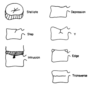
Various types of endplate fracture have been found to occur in the lumbar spine (figure 1). In some instances there can be intrusion of disc material into the trabecular bone called intraosseous herniation. This defect can become calcified in what is called a Schmorl's node. In adolescents, endplate failure may occur differently with fracture of the posterior edge that runs from the endplate down to the cartilage growth plate. Endplate fractures are not always seen on plain films. MRI is more accurate and this modality can also show Modic changes which are reactions to the endplate fracture.
Osteoporotic and Elderly Vertebra

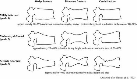
Osteoporotic vertebra display different patterns of failure: anterior wedge fractures, biconcave fractures, and crush fractures (figure 2).
Anterior wedge fractures are where there is collapse of the cortex of the vertebral body plus collapse of the underlying trabecular bone leading to a wedge shape. It is thought that with osteoporosis there is relative loss of thickness of the anterior trabeculae compared to the posterior trabeculae. There is also relative loss of the horizontal compared to the vertebral trabeculae, which reduces resistance to tension. Furthermore, the endplate is weaker anteriorly than posteriorly. With intervertebral height reduction, more of the compressive load is taken up by the posterior column, and the anterior column is "stress-shielded" in the neutral and extended positions. However in the flexed position the anterior vertebral body is severely loaded in the degenerative disc. The elderly may spend less time in flexion leading to a reduced mechanical stimulus for hypertrophic adaptation. This combined with a reduced ability for the endplate to adapt (e.g. due to menopause), may explain why fractures occur in the anterior vertebral body and why flexion movements often precipitate it.[3]
Biconcave fractures are also common in the elderly. In this type of fracture both endplates have collapsed inwards. The endplates are often smooth which suggests repeated microfractures having occurred. In younger spines compression failure usually leads to more discontinuous endplate damage. Back to the elderly, the discs may not have an overall reduced height even though degeneration has occurred. This is because the nucleus has less resistance with pushing into the weakened vertebra. The annulus fibrosus may be reduced in thickness, however, and this may be a better sign of intervertebral disc integrity than overall height or nucleus height.
Crush fractures are thought to be due to a sudden compressive force acting on the anterior and posterior vertebral cortices at the same time. In younger spines this type of trauma can cause burst fractures which have a similar appearance.
References
- Stellate fracture of the endplate (stellate): two or more cracks running from the centre towards the periphery of the endplate;
- Step-like fracture of the endplate (step): a crack in the endplate with a distinctly visible step of l-4 mm height;
- Endplate depression (depression): endplate deformed into a bowl shape, the trabecular bone below the endplate fractured;
- Transverse fracture (transverse): transverse fracture through the whole vertebra, usually in its midplane;
- Edge fracture (ed ventral or dorsal): wedge-like fracture of the edge of the vertebral body;
- Y-fracture (y-fract): combination of a depression of the endplate with a fracture line approximately in the axis of the vertebra;
- Disc intrusion (intr): intrusion of disc material into the trabecular bone observed in combination with some of the above fracture types
- ↑ Brinckmann P, Biggemann M, Hilweg D. Prediction of the compressive strength of human lumbar vertebrae. Clin Biomech (Bristol, Avon). 1989;4 Suppl 2:iii-27. doi: 10.1016/0268-0033(89)90071-5. PMID: 23906213.
- ↑ Brinckmann P, Biggemann M, Hilweg D. Prediction of the compressive strength of human lumbar vertebrae. Clin Biomech (Bristol, Avon). 1989;4 Suppl 2:iii-27. doi: 10.1016/0268-0033(89)90071-5. PMID: 23906213.
- ↑ Pollintine P, Dolan P, Tobias JH, Adams MA. Intervertebral disc degeneration can lead to "stress-shielding" of the anterior vertebral body: a cause of osteoporotic vertebral fracture? Spine (Phila Pa 1976). 2004 Apr 1;29(7):774-82. doi: 10.1097/01.brs.0000119401.23006.d2. PMID: 15087801.

