File:Blood nerve barrier.jpg

Original file (1,977 × 1,508 pixels, file size: 263 KB, MIME type: image/jpeg)
Summary
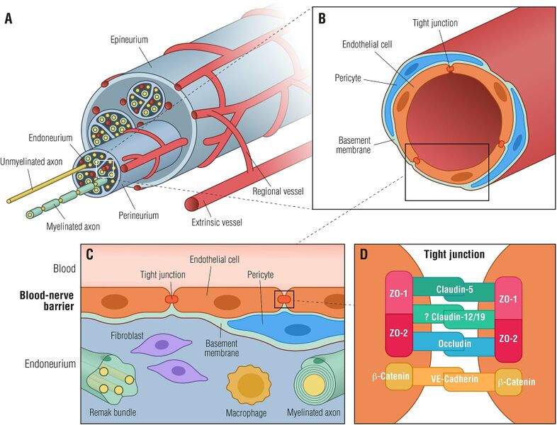
Figure 1. Blood-nerve barrier. (A) Transverse view of a peripheral nerve ensheathed by epineurial collagen fibrils (epineurium) and blood vessels. Individual nerve fascicles consisting of unmyelinated and myelinated axons as well as small blood vessels are ensheathed by the perineurium, forming the endoneurial microenvironment. (B) Individual endoneurial blood vessel surrounded by endothelial cells, pericytes and the basement membrane. (C) Cellular structure of the blood-nerve barrier, formed by endothelial cells, that are connected by tight junctions, pericytes and the basement membrane. The barrier is exposed to cells and molecules circulating in the blood, protecting constituents of the endoneurium (Remak bundles, myelinated axons, resident macrophages and fibroblasts) from toxic factors. (D) Endothelial cells are tightly interconnected by tight junctions and adherens junctions forming a restrictive intercellular barrier. Zona occludens-1 and -2 (ZO-1, ZO-2) interact with claudin-5, occludin and likely with claudin-12/19 forming tight junctions. β-catenin forms in conjunction with VE-cadherin adherens junctions.[1]
Licencing
![]()
This work is licensed under a Creative Commons Attribution 4.0 International License.
- ↑ Richner, Mette et al. “Functional and Structural Changes of the Blood-Nerve-Barrier in Diabetic Neuropathy.” Frontiers in neuroscience vol. 12 1038. 14 Jan. 2019, doi:10.3389/fnins.2018.01038
File history
Click on a date/time to view the file as it appeared at that time.
| Date/Time | Thumbnail | Dimensions | User | Comment | |
|---|---|---|---|---|---|
| current | 03:08, 6 December 2021 | | 1,977 × 1,508 (263 KB) | Jeremy (talk | contribs) | Figure 1. Blood-nerve barrier. (A) Transverse view of a peripheral nerve ensheathed by epineurial collagen fibrils (epineurium) and blood vessels. Individual nerve fascicles consisting of unmyelinated and myelinated axons as well as small blood vessels are ensheathed by the perineurium, forming the endoneurial microenvironment. (B) Individual endoneurial blood vessel surrounded by endothelial cells, pericytes and the basement membrane. (C) Cellular structure of the blood-nerve barrier, formed... |
You cannot overwrite this file.
File usage
There are no pages that use this file.

