File:Clavicle shortening.jpg
From WikiMSK

Size of this preview: 800 × 166 pixels. Other resolution: 834 × 173 pixels.
Original file (834 × 173 pixels, file size: 25 KB, MIME type: image/jpeg)
Summary
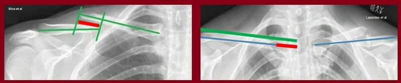
Figure 4: Shortening of the clavicle can be assessed by either of the following methods. On the left, the overlap between the ends of the medial and lateral fragments is measured directly; this is shown in red. At right, the entire medial-to-lateral length of both the injured and the normal clavicle are measured. As shown, the blue line represents the length of the shortened clavicle, the green line shows the length of the normal one, and the red line represents the difference. (from https://bmcmusculoskeletdisord.biomedcentral.com/articles/10.1186/s12891-017-1881-x)
Licencing
![]()
This work is licensed under the Creative Commons Attribution-NonCommercial-ShareAlike License.
File history
Click on a date/time to view the file as it appeared at that time.
| Date/Time | Thumbnail | Dimensions | User | Comment | |
|---|---|---|---|---|---|
| current | 05:58, 8 March 2022 | 834 × 173 (25 KB) | Jeremy (talk | contribs) | Figure 4: Shortening of the clavicle can be assessed by either of the following methods. On the left, the overlap between the ends of the medial and lateral fragments is measured directly; this is shown in red. At right, the entire medial-to-lateral length of both the injured and the normal clavicle are measured. As shown, the blue line represents the length of the shortened clavicle, the green line shows the length of the normal one, and the red line represents the difference. (from https://... |
You cannot overwrite this file.
File usage
The following page uses this file:

