File:Costameres.jpg
Costameres.jpg (363 × 482 pixels, file size: 76 KB, MIME type: image/jpeg)
Summary
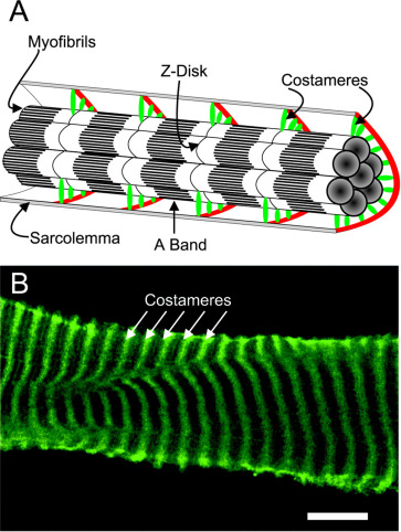
Cellular location of costameres in striated muscle. Shown in A is a schematic diagram illustrating costameres as circumferential elements that physically couple peripheral myofibrils to the sarcolemma in periodic register with the Z-disk. The protein composition of costameres is shown in expanded form in Fig. 2. Shown in B is an inside-out sarcolemma that was mechanically peeled from a single myofibre and stained with antibodies to γ-actin to reveal the costameric cytoskeleton. Bar, 10 μm. B is reproduced from Ref. 17 by copyright permission of The Rockefeller University Press
From Ervasti JM. Costameres: the Achilles' heel of Herculean muscle. J Biol Chem. 2003 Apr 18;278(16):13591-4. doi: 10.1074/jbc.R200021200. Epub 2003 Jan 29. PMID: 12556452.
Licencing
![]()
This work is licensed under a Creative Commons Attribution 4.0 International License.
File history
Click on a date/time to view the file as it appeared at that time.
| Date/Time | Thumbnail | Dimensions | User | Comment | |
|---|---|---|---|---|---|
| current | 15:29, 10 August 2021 | | 363 × 482 (76 KB) | Jeremy (talk | contribs) | From Ervasti JM. Costameres: the Achilles' heel of Herculean muscle. J Biol Chem. 2003 Apr 18;278(16):13591-4. doi: 10.1074/jbc.R200021200. Epub 2003 Jan 29. PMID: 12556452. |
You cannot overwrite this file.
File usage
The following page uses this file:

