File:Cutting cone simplified.png

Original file (850 × 471 pixels, file size: 36 KB, MIME type: image/png)
Summary
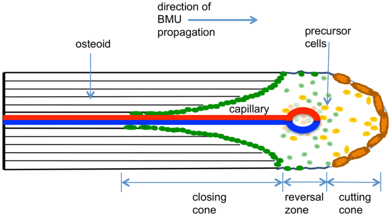
Idealized structure of cortical BMU in longitudinal section, showing cutting cone, reversal zone and closing cone. Cells of the osteoclast lineage are shown in light to dark orange while cells of the osteoblastic lineage are shown as light to dark green. A capillary is shown at the centre of the BMU within a Haversian canal.
Smith DW, Gardiner BS, Dunstan C. Bone balance within a cortical BMU: local controls of bone resorption and formation. PLoS One. 2012;7(7):e40268. doi: 10.1371/journal.pone.0040268. Epub 2012 Jul 23. PMID: 22844401; PMCID: PMC3402480.
Licencing
![]()
This work is licensed under a Creative Commons Attribution 4.0 International License.
File history
Click on a date/time to view the file as it appeared at that time.
| Date/Time | Thumbnail | Dimensions | User | Comment | |
|---|---|---|---|---|---|
| current | 11:30, 1 August 2021 | | 850 × 471 (36 KB) | Jeremy (talk | contribs) | Idealized structure of cortical BMU in longitudinal section, showing cutting cone, reversal zone and closing cone. Cells of the osteoclast lineage are shown in light to dark orange while cells of the osteoblastic lineage are shown as light to dark green. A capillary is shown at the centre of the BMU within a Haversian canal. Smith DW, Gardiner BS, Dunstan C. Bone balance within a cortical BMU: local controls of bone resorption and formation. PLoS One. 2012;7(7):e40268. doi: 10.1371/journal.p... |
You cannot overwrite this file.
File usage
The following page uses this file:

