File:Femur bone segments.jpg
From WikiMSK
No higher resolution available.
Femur_bone_segments.jpg (512 × 586 pixels, file size: 28 KB, MIME type: image/jpeg)
Summary
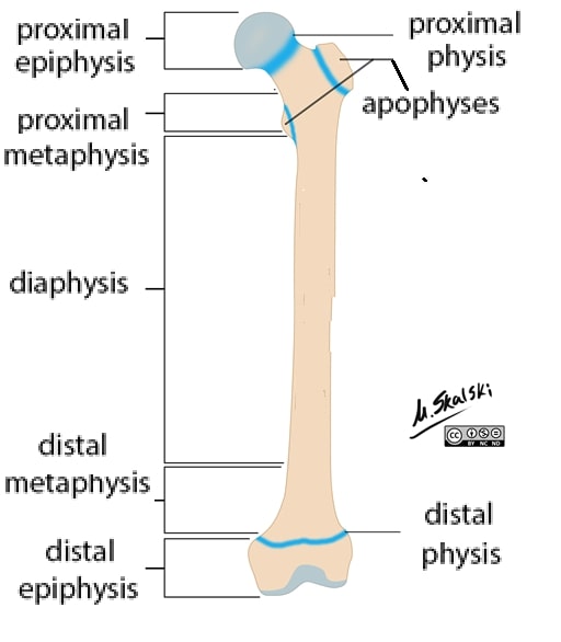
Figure 1: Bone segments of the femur. As the figure makes clear, in a long bone there is only one diaphysis, but there can be physes and associated regions at both ends. Also shown are the apophyses, the growth plates of the secondary growth centers where tendons attach [In the proximal femur, these will become the trochanters.] (Modified from Dr Matt Skalski, Radiopaedia.org. From the case https://radiopaedia.org/cases/29729)
Licencing
![]()
This work is licensed under the Creative Commons Attribution-NonCommercial-ShareAlike License.
File history
Click on a date/time to view the file as it appeared at that time.
| Date/Time | Thumbnail | Dimensions | User | Comment | |
|---|---|---|---|---|---|
| current | 19:30, 8 March 2022 | | 512 × 586 (28 KB) | Jeremy (talk | contribs) | Figure 1: Bone segments of the femur. As the figure makes clear, in a long bone there is only one diaphysis, but there can be physes and associated regions at both ends. Also shown are the apophyses, the growth plates of the secondary growth centers where tendons attach [In the proximal femur, these will become the trochanters.] (Modified from Dr Matt Skalski, Radiopaedia.org. From the case https://radiopaedia.org/cases/29729) |
You cannot overwrite this file.
File usage
The following page uses this file:

