File:Foot and Ankle Radiograph Normal.jpg
From WikiMSK

Size of this preview: 510 × 600 pixels. Other resolution: 647 × 761 pixels.
Original file (647 × 761 pixels, file size: 59 KB, MIME type: image/jpeg)
Summary
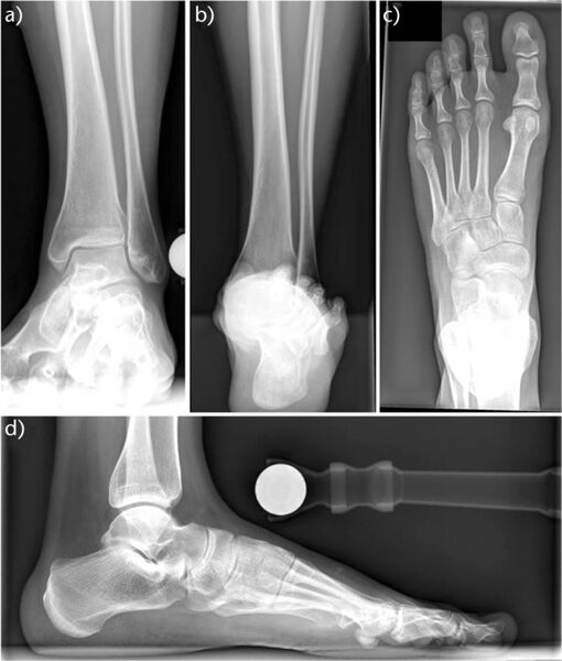
Plain weight-bearing radiographs of a healthy foot and ankle: a) anteroposterior view; b) hindfoot alignment view; c) dorso-plantar view; d) lateral view.
Krähenbühl, Nicola et al. “The subtalar joint: A complex mechanism.” EFORT open reviews vol. 2,7 309-316. 6 Jul. 2017, doi:10.1302/2058-5241.2.160050
Licencing
![]()
This work is licensed under the Creative Commons Attribution-NonCommercial 4.0 License.
File history
Click on a date/time to view the file as it appeared at that time.
| Date/Time | Thumbnail | Dimensions | User | Comment | |
|---|---|---|---|---|---|
| current | 13:51, 17 July 2021 | | 647 × 761 (59 KB) | Jeremy (talk | contribs) | Plain weight-bearing radiographs of a healthy foot and ankle: a) anteroposterior view; b) hindfoot alignment view; c) dorso-plantar view; d) lateral view. Krähenbühl, Nicola et al. “The subtalar joint: A complex mechanism.” EFORT open reviews vol. 2,7 309-316. 6 Jul. 2017, doi:10.1302/2058-5241.2.160050 |
You cannot overwrite this file.
File usage
The following page uses this file:

