File:GON and TON ultrasound.png
Original file (950 × 494 pixels, file size: 480 KB, MIME type: image/png)
Summary
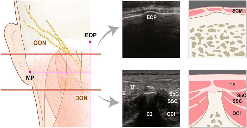
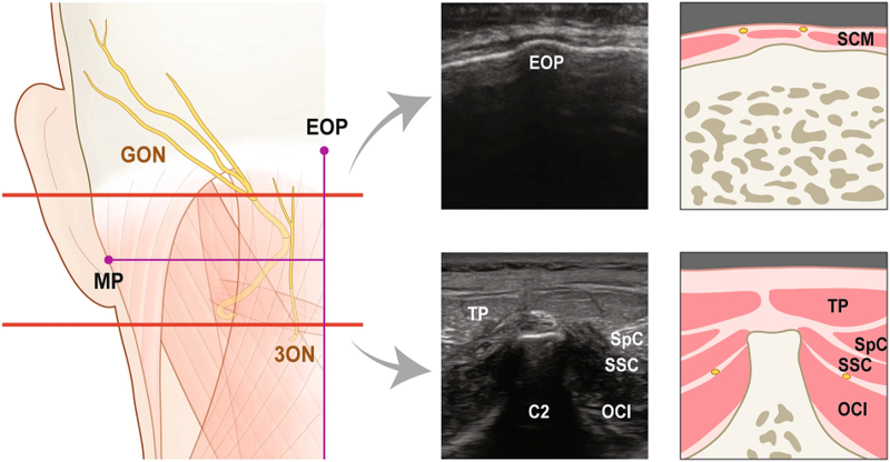
The position of greater occipital nerve (GON) and third occipital nerve (3ON) in cadaveric observation (left illustration) and the corresponding ultrasound images (right images). Upper red line runs through the emergence of GON from the trapezius (TP) and lower one through the spinous process of the axis (C2). Each ultrasound images and their illustrations corresponds to sections through upper and lower red lines respectively. sternocleidomastoid muscle, SCM; splenius capitis, SpC; semispinalis capitis, SSC; obliquus capitis inferior, OCI. Kim et al.. Stereotactic topography of the greater and third occipital nerves and its clinical implication. Scientific reports 2018. 8:870. PMID: 29343808. DOI. Full Text.
Licencing
![]()
This work is licensed under a Creative Commons Attribution 4.0 International License.
File history
Click on a date/time to view the file as it appeared at that time.
| Date/Time | Thumbnail | Dimensions | User | Comment | |
|---|---|---|---|---|---|
| current | 10:58, 11 August 2020 | | 950 × 494 (480 KB) | Jeremy (talk | contribs) | The position of greater occipital nerve (GON) and third occipital nerve (3ON) in cadaveric observation (left illustration) and the corresponding ultrasound images (right images). Upper red line runs through the emergence of GON from the trapezius (TP) and lower one through the spinous process of the axis (C2). Each ultrasound images and their illustrations corresponds to sections through upper and lower red lines respectively. sternocleidomastoid muscle, SCM; splenius capitis, SpC; semispinal... |
You cannot overwrite this file.
File usage
The following 2 pages use this file:

