File:Glenohumeral joint capsular structures.jpg
From WikiMSK

Size of this preview: 441 × 599 pixels. Other resolution: 717 × 974 pixels.
Original file (717 × 974 pixels, file size: 94 KB, MIME type: image/jpeg)
Summary
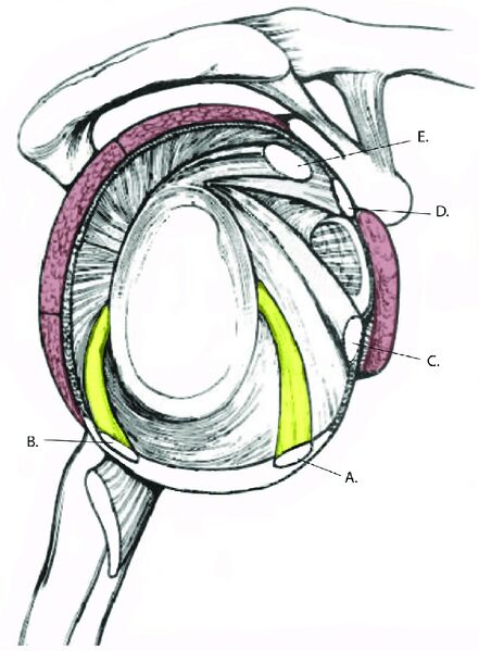
Anatomic diagram of the glenohumeral joint capsular structures. A: Anterior band of the inferior glenohuemral ligament (IGL); B: posterior band of the IGL. C: middle glenohumeral ligament; D: superior glenohumeral ligament. E: long head of biceps tendon. The structures in red are the muscles of the rotator cuff.
From https://www.arthroscopytechniques.org/article/S2212-6287(20)30212-7/fulltext
Licencing
![]()
This work is licensed under a Creative Commons Attribution 4.0 International License.
File history
Click on a date/time to view the file as it appeared at that time.
| Date/Time | Thumbnail | Dimensions | User | Comment | |
|---|---|---|---|---|---|
| current | 13:13, 19 August 2021 | | 717 × 974 (94 KB) | Jeremy (talk | contribs) | Anatomic diagram of the glenohumeral joint capsular structures. A: Anterior band of the inferior glenohuemral ligament (IGL); B: posterior band of the IGL. C: middle glenohumeral ligament; D: superior glenohumeral ligament. E: long head of biceps tendon. The structures in red are the muscles of the rotator cuff. From https://www.arthroscopytechniques.org/article/S2212-6287(20)30212-7/fulltext |
You cannot overwrite this file.
File usage
The following page uses this file:

