File:Gluteus medius MRI high grade partial thickness tear.jpg
Gluteus_medius_MRI_high_grade_partial_thickness_tear.jpg (688 × 368 pixels, file size: 85 KB, MIME type: image/jpeg)
Summary
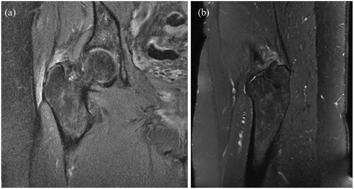
(a) Coronal fat suppressed proton density and (b) sagittal T2-weighted sequences on MRI of the right hip showing a high-grade partial tear of the gluteus medius and minimus tendons with tendinosis and underlying trochanteric bursitis. The patient consented for publication of this imaging.
Pianka MA, Serino J, DeFroda SF, Bodendorfer BM. Greater trochanteric pain syndrome: Evaluation and management of a wide spectrum of pathology. SAGE Open Med. 2021 Jun 3;9:20503121211022582. doi: 10.1177/20503121211022582. PMID: 34158938; PMCID: PMC8182177.
Licencing
![]()
This work is licensed under the Creative Commons Attribution-NonCommercial 4.0 License.
File history
Click on a date/time to view the file as it appeared at that time.
| Date/Time | Thumbnail | Dimensions | User | Comment | |
|---|---|---|---|---|---|
| current | 20:03, 11 April 2022 | | 688 × 368 (85 KB) | Jeremy (talk | contribs) | (a) Coronal fat suppressed proton density and (b) sagittal T2-weighted sequences on MRI of the right hip showing a high-grade partial tear of the gluteus medius and minimus tendons with tendinosis and underlying trochanteric bursitis. The patient consented for publication of this imaging. Pianka MA, Serino J, DeFroda SF, Bodendorfer BM. Greater trochanteric pain syndrome: Evaluation and management of a wide spectrum of pathology. SAGE Open Med. 2021 Jun 3;9:20503121211022582. doi: 10.1177/20... |
You cannot overwrite this file.
File usage
The following page uses this file:

