File:Hindfoot alignment radiograph.jpg
Original file (634 × 1,100 pixels, file size: 113 KB, MIME type: image/jpeg)
Summary
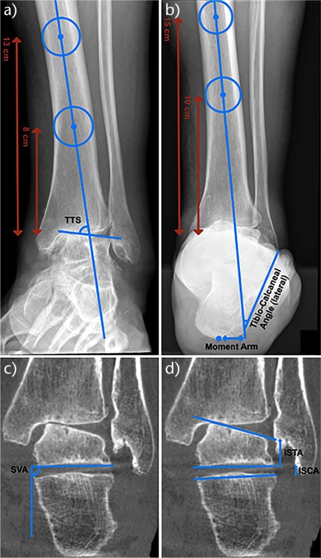
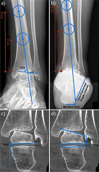
Hindfoot alignment measurements using plain weight-bearing radiographs and weight-bearing CT scans: a) anteroposterior view, tibiotalar surface angle (TTS) is used to measure inclination of the talus in relation to the tibial axis; b) hindfoot alignment view, the moment arm of the calcaneus and lateral tibio-calcaneal angle are used to assess the hindfoot alignment; c) weight-bearing CT scan, subtalar vertical angle (SVA) is used to measure the subtalar joint axis relative to the ground; d) weight-bearing CT scan, inftal-subtal angle (ISTA) is used to assess the talar morphology and inftal-supcal angle (ISCA) to determine the inclination of the calcaneus in relation to the talus.
Krähenbühl, Nicola et al. “The subtalar joint: A complex mechanism.” EFORT open reviews vol. 2,7 309-316. 6 Jul. 2017, doi:10.1302/2058-5241.2.160050
Licencing
![]()
This work is licensed under the Creative Commons Attribution-NonCommercial 4.0 License.
File history
Click on a date/time to view the file as it appeared at that time.
| Date/Time | Thumbnail | Dimensions | User | Comment | |
|---|---|---|---|---|---|
| current | 13:53, 17 July 2021 | | 634 × 1,100 (113 KB) | Jeremy (talk | contribs) | Hindfoot alignment measurements using plain weight-bearing radiographs and weight-bearing CT scans: a) anteroposterior view, tibiotalar surface angle (TTS) is used to measure inclination of the talus in relation to the tibial axis; b) hindfoot alignment view, the moment arm of the calcaneus and lateral tibio-calcaneal angle are used to assess the hindfoot alignment; c) weight-bearing CT scan, subtalar vertical angle (SVA) is used to measure the subtalar joint axis relative to the ground; d) w... |
You cannot overwrite this file.
File usage
The following page uses this file:

