File:Hip joint injection anterolateral approach ultrasound.jpg
From WikiMSK
Size of this preview: 613 × 600 pixels. Other resolution: 640 × 626 pixels.
Original file (640 × 626 pixels, file size: 56 KB, MIME type: image/jpeg)
Summary
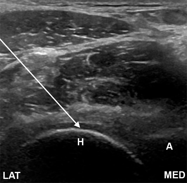
Transverse image of the hip joint. The needle is introduced from a lateral and anterior approach, to rest on the femoral head (arrow). A, acetabulum; H, femoral head; N, femoral neck; LAT, lateral; MED, medial.
Yeap & Robinson. Ultrasound Diagnostic and Therapeutic Injections of the Hip and Groin. Journal of the Belgian Society of Radiology 2017. 101:6. PMID: 30498802. DOI. Full Text.
Licencing
![]()
This work is licensed under a Creative Commons Attribution 4.0 International License.
File history
Click on a date/time to view the file as it appeared at that time.
| Date/Time | Thumbnail | Dimensions | User | Comment | |
|---|---|---|---|---|---|
| current | 14:59, 21 June 2021 | | 640 × 626 (56 KB) | Jeremy (talk | contribs) | Transverse image of the hip joint. The needle is introduced from a lateral and anterior approach, to rest on the femoral head (arrow). A, acetabulum; H, femoral head; N, femoral neck; LAT, lateral; MED, medial. {{#pmid:30498802}} |
You cannot overwrite this file.
File usage
The following page uses this file:

