File:Lateral hip illustration and cadaver.jpg
From WikiMSK
No higher resolution available.
Lateral_hip_illustration_and_cadaver.jpg (624 × 360 pixels, file size: 48 KB, MIME type: image/jpeg)
Summary
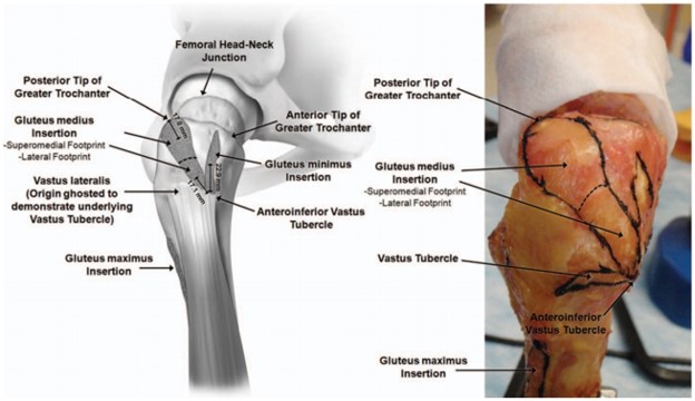
(Left) Illustration and (right) photograph of lateral view of a right hip looking medially at the footprint insertions of the greater trochanter. The footprints of the gluteus medius, gluteus minimus, and vastus lateralis with respect to the vastus tubercle are depicted (Philippon et al. [34]).
From J Hip Preserv Surg, Volume 6, Issue 4, December 2019, Pages 398–405, https://doi.org/10.1093/jhps/hnz046
Licencing
![]()
This work is licensed under the Creative Commons Attribution-NonCommercial 4.0 License.
File history
Click on a date/time to view the file as it appeared at that time.
| Date/Time | Thumbnail | Dimensions | User | Comment | |
|---|---|---|---|---|---|
| current | 17:54, 11 April 2022 | | 624 × 360 (48 KB) | Jeremy (talk | contribs) | (Left) Illustration and (right) photograph of lateral view of a right hip looking medially at the footprint insertions of the greater trochanter. The footprints of the gluteus medius, gluteus minimus, and vastus lateralis with respect to the vastus tubercle are depicted (Philippon et al. [34]). From J Hip Preserv Surg, Volume 6, Issue 4, December 2019, Pages 398–405, https://doi.org/10.1093/jhps/hnz046 |
You cannot overwrite this file.
File usage
The following page uses this file:

