File:Medial hamstring reflex.PNG
Medial_hamstring_reflex.PNG (502 × 369 pixels, file size: 290 KB, MIME type: image/png)
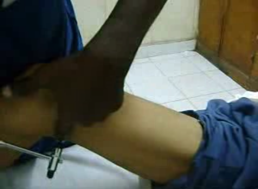
"With the patient in the Supine position, hip slightly flexed, externally rotated and abducted, the ipsilateral knee is semi flexed and supported by one of the examiner's hands. The reflex muscle contraction was elicited by striking the index finger of the supporting hand placed on the medial hamstring tendon (tendons of semitendinosus, semimembranosus) just above the knee join (postero-medially) with a reflex hammer. The normal response is contraction of the medial mass of hamstring muscles [Video 1]. Knee flexion is hardly observed because of friction between heel and couch. The action of the semimembranosus and semitendinosus muscles is to extend the hip joint, flex and internally rotate the knee. The MHR is mediated by the tibial portion of the sciatic nerve, primarily by the L5 nerve root and is the only deep tendon reflex useful in the evaluation of suspected L5 radiculopathy. It is shown that in the presence of symmetrically active gastrocsoleus reflexes, asymmetry of the hamstring reflexes indicates an L5 root lesion."[1]
File history
Click on a date/time to view the file as it appeared at that time.
| Date/Time | Thumbnail | Dimensions | User | Comment | |
|---|---|---|---|---|---|
| current | 19:04, 19 October 2020 | | 502 × 369 (290 KB) | Jeremy (talk | contribs) |
You cannot overwrite this file.
File usage
There are no pages that use this file.

