File:SFN biopsies.jpg
SFN_biopsies.jpg (700 × 304 pixels, file size: 59 KB, MIME type: image/jpeg)
Summary
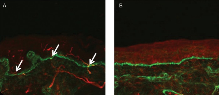
Confocal images of skin biopsies taken from the legs of a control subject (A) and a patient with small fibre neuropathy secondary to HIV (B) showing PGP 9.5-immunoreactive fibres (red) and the basement membrane (labelled with type IV collagen fibres, green). Nerve fibres positive for PGP 9.5 (white arrows) are counted as they cross the dermal–epidermal junction. The intra-epidermal nerve fibres are absent in the patient with HIV (B) consistent with the diagnosis of small fibre neuropathy. Scale bar: 50 µm
Themistocleous AC, Ramirez JD, Serra J, Bennett DL. The clinical approach to small fibre neuropathy and painful channelopathy. Pract Neurol. 2014 Dec;14(6):368-79. doi: 10.1136/practneurol-2013-000758. Epub 2014 Apr 28. PMID: 24778270; PMCID: PMC4251302.
Licencing
![]()
This work is licensed under a Creative Commons Attribution 4.0 International License.
File history
Click on a date/time to view the file as it appeared at that time.
| Date/Time | Thumbnail | Dimensions | User | Comment | |
|---|---|---|---|---|---|
| current | 19:18, 15 March 2023 | | 700 × 304 (59 KB) | Jeremy (talk | contribs) | Confocal images of skin biopsies taken from the legs of a control subject (A) and a patient with small fibre neuropathy secondary to HIV (B) showing PGP 9.5-immunoreactive fibres (red) and the basement membrane (labelled with type IV collagen fibres, green). Nerve fibres positive for PGP 9.5 (white arrows) are counted as they cross the dermal–epidermal junction. The intra-epidermal nerve fibres are absent in the patient with HIV (B) consistent with the diagnosis of small fibre neuropathy. Sca... |
You cannot overwrite this file.
File usage
The following 4 pages use this file:

