File:SIJ fluoroscopy AP technique.jpg
SIJ_fluoroscopy_AP_technique.jpg (669 × 295 pixels, file size: 40 KB, MIME type: image/jpeg)
Summary
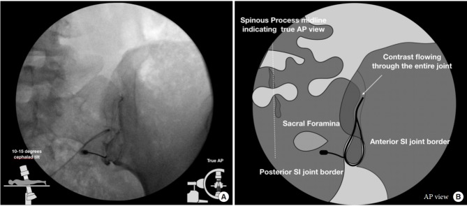
Anteroposterior technique fluoroscopic view (A) and graphical illustration (B). In the anteriorposterior (AP) approach, image is taken with 5–15 degree cephalad tilt from the vertical of the fluoroscopy machine reveals a joint with 2 separately visible anterior and posterior joint lines. The anterior and posterior parts of the sacroiliac (SI) joint were delineated as lateral and medial joint spaces, respectively.
From Chauhan G, Hehar P, Loomba V, Upadhyay A. A Randomized Controlled Trial of Fluoroscopically-Guided Sacroiliac Joint Injections: A Comparison of the Posteroanterior and Classical Oblique Techniques. Neurospine. 2019 Jun;16(2):317-324. doi: 10.14245/ns.1836122.061. Epub 2018 Oct 7. PMID: 30531656; PMCID: PMC6603830.
Licencing
![]()
This work is licensed under the Creative Commons Attribution-NonCommercial 4.0 License.
File history
Click on a date/time to view the file as it appeared at that time.
| Date/Time | Thumbnail | Dimensions | User | Comment | |
|---|---|---|---|---|---|
| current | 08:51, 15 April 2022 | | 669 × 295 (40 KB) | Jeremy (talk | contribs) | Anteroposterior technique fluoroscopic view (A) and graphical illustration (B). In the anteriorposterior (AP) approach, image is taken with 5–15 degree cephalad tilt from the vertical of the fluoroscopy machine reveals a joint with 2 separately visible anterior and posterior joint lines. The anterior and posterior parts of the sacroiliac (SI) joint were delineated as lateral and medial joint spaces, respectively. From Chauhan G, Hehar P, Loomba V, Upadhyay A. A Randomized Controlled Trial of... |
You cannot overwrite this file.
File usage
The following page uses this file:

