File:SIJ fluoroscopy oblique technique.jpg
SIJ_fluoroscopy_oblique_technique.jpg (671 × 296 pixels, file size: 41 KB, MIME type: image/jpeg)
Summary
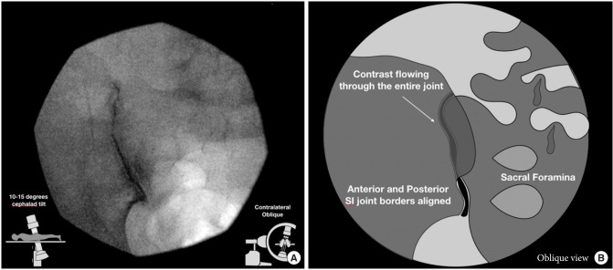
Oblique technique fluoroscopic view (A) and graphical illustration (B). In the oblique approach, the C-arm is rotated in a contralateral manner until the 2 joint lines become superimposed. Then one would target the inferior segment of this superimposed image, as the superior sacroiliac (SI) joint space is composed of interosseous ligaments.
From Chauhan G, Hehar P, Loomba V, Upadhyay A. A Randomized Controlled Trial of Fluoroscopically-Guided Sacroiliac Joint Injections: A Comparison of the Posteroanterior and Classical Oblique Techniques. Neurospine. 2019 Jun;16(2):317-324. doi: 10.14245/ns.1836122.061. Epub 2018 Oct 7. PMID: 30531656; PMCID: PMC6603830.
Licencing
![]()
This work is licensed under the Creative Commons Attribution-NonCommercial 4.0 License.
File history
Click on a date/time to view the file as it appeared at that time.
| Date/Time | Thumbnail | Dimensions | User | Comment | |
|---|---|---|---|---|---|
| current | 08:51, 15 April 2022 | | 671 × 296 (41 KB) | Jeremy (talk | contribs) | Oblique technique fluoroscopic view (A) and graphical illustration (B). In the oblique approach, the C-arm is rotated in a contralateral manner until the 2 joint lines become superimposed. Then one would target the inferior segment of this superimposed image, as the superior sacroiliac (SI) joint space is composed of interosseous ligaments. From Chauhan G, Hehar P, Loomba V, Upadhyay A. A Randomized Controlled Trial of Fluoroscopically-Guided Sacroiliac Joint Injections: A Comparison of the... |
You cannot overwrite this file.
File usage
The following page uses this file:

