File:Sagittal band tear ultrasound.jpg
From WikiMSK
Size of this preview: 800 × 314 pixels. Other resolution: 1,026 × 403 pixels.
Original file (1,026 × 403 pixels, file size: 82 KB, MIME type: image/jpeg)
Summary
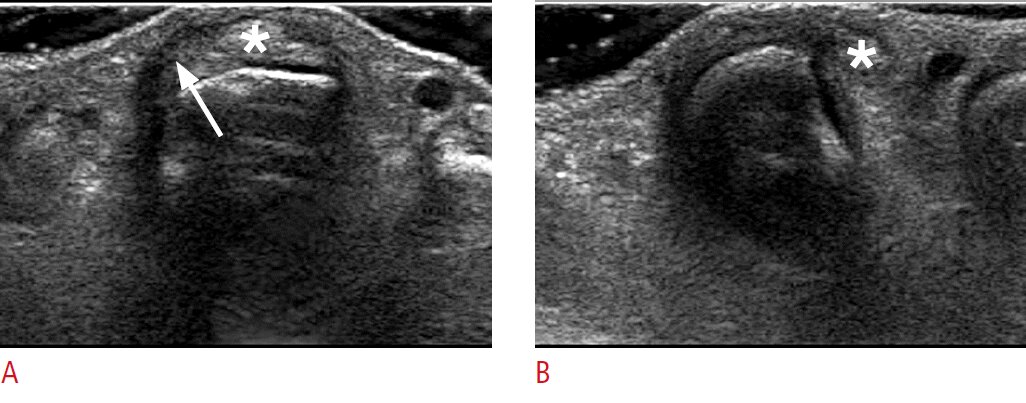
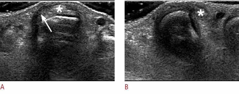
Sagittal band tear in a 43-year-old woman. A. A transverse sonogram of the third metacarpophalangeal joint when the finger is extended shows an abnormal radial sagittal band with irregularity and hypoechogenicity (arrow). The extensor tendon (asterisk) is positioned normally. B. A dynamic examination obtained in the transverse plane during finger flexion shows dislocation of the extensor tendon (asterisk).[1]
Licencing
![]()
This work is licensed under the Creative Commons Attribution-NonCommercial 4.0 License.
- ↑ Lee SA, Kim BH, Kim SJ, Kim JN, Park SY, Choi K. Current status of ultrasonography of the finger. Ultrasonography. 2016 Apr;35(2):110-23. doi: 10.14366/usg.15051. Epub 2015 Nov 24. PMID: 26753604; PMCID: PMC4825212.
File history
Click on a date/time to view the file as it appeared at that time.
| Date/Time | Thumbnail | Dimensions | User | Comment | |
|---|---|---|---|---|---|
| current | 12:30, 6 February 2022 | 1,026 × 403 (82 KB) | Jeremy (talk | contribs) | Sagittal band tear in a 43-year-old woman. A. A transverse sonogram of the third metacarpophalangeal joint when the finger is extended shows an abnormal radial sagittal band with irregularity and hypoechogenicity (arrow). The extensor tendon (asterisk) is positioned normally. B. A dynamic examination obtained in the transverse plane during finger flexion shows dislocation of the extensor tendon (asterisk).<ref>Lee SA, Kim BH, Kim SJ, Kim JN, Park SY, Choi K. Current status of ultrasonography... |
You cannot overwrite this file.
File usage
The following page uses this file:

