File:Tendinopathy histology.jpg
Tendinopathy_histology.jpg (358 × 457 pixels, file size: 66 KB, MIME type: image/jpeg)
Summary
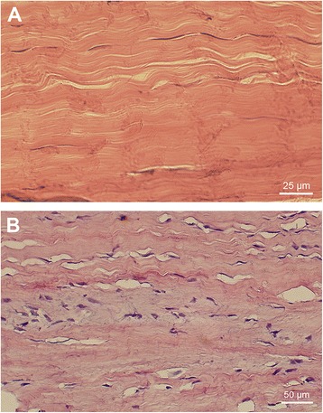
Histological differences between normal and tendinosis tendon. The normal tendon shows organised collagen fibres and a sparse amount of tendon cells, tightly packed between the collagen bundles (A). In tendinosis (B), the tendon structure gets disorganised, the tenocytes change morphology and proliferate.
From Christensen J, Alfredson H, Andersson G. Protease-activated receptors in the Achilles tendon-a potential explanation for the excessive pain signalling in tendinopathy. Mol Pain. 2015 Mar 17;11:13. doi: 10.1186/s12990-015-0007-4. PMID: 25880199; PMCID: PMC4369088.
Licencing
![]()
This work is licensed under a Creative Commons Attribution 4.0 International License.
File history
Click on a date/time to view the file as it appeared at that time.
| Date/Time | Thumbnail | Dimensions | User | Comment | |
|---|---|---|---|---|---|
| current | 23:00, 11 August 2021 | | 358 × 457 (66 KB) | Jeremy (talk | contribs) | Histological differences between normal and tendinosis tendon. The normal tendon shows organised collagen fibres and a sparse amount of tendon cells, tightly packed between the collagen bundles (A). In tendinosis (B), the tendon structure gets disorganised, the tenocytes change morphology and proliferate. From Christensen J, Alfredson H, Andersson G. Protease-activated receptors in the Achilles tendon-a potential explanation for the excessive pain signalling in tendinopathy. Mol Pain. 2015 M... |
You cannot overwrite this file.
File usage
The following page uses this file:

