Uncategorised files
From WikiMSK
Showing below up to 50 results in range #501 to #550.
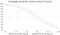 Lidocaine vs bupivicaine inactive form percentage vs ph.png 1,718 × 1,020; 108 KB
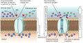 Ligand-gated-channels.jpg 1,176 × 611; 177 KB
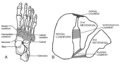 Lisfranc joint.png 1,168 × 614; 256 KB
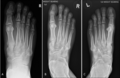 Lisfranc xray.PNG 589 × 385; 251 KB
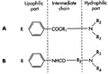 Local anaesthetic structure.png 585 × 404; 36 KB
 Low Back Pain - Knezevic 2021.pdf ; 667 KB
- Low back pain PROMs.pdf ; 1.16 MB
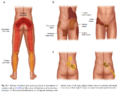
 Low back pain somatic and radicular features.PNG 793 × 748; 141 KB
 Low back pain taxonomy.jpg 826 × 1,529; 120 KB
 Lowback.png 128 × 128; 7 KB
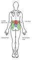
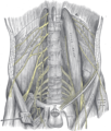
 Lumbar-Plexus-Anatomy-1.jpg 600 × 526; 101 KB
 Lumbar-medial-branch-nerve-blocks.jpg 717 × 806; 55 KB
 Lumbar-medial-branch-nerve-blocks2.jpg 637 × 803; 61 KB
 Lumbar-neuroforaminal-stenosis-normal.jpg 686 × 686; 52 KB
 Lumbar-plexus-and-its-branches.png 508 × 600; 95 KB
 Lumbar Facet pain.PNG 783 × 613; 441 KB
 Lumbar interspinous oedema hierarchy.png 1,006 × 330; 12 KB
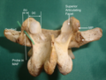
 Lumbar mamillo-accessory ligament.png 467 × 253; 98 KB
 Lumbar mamillo-accessory ligament posterior view.png 352 × 266; 203 KB
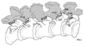
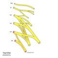
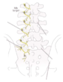 Lumbar medial branches.png 524 × 682; 248 KB
 Lumbosacral trunk.jpeg 630 × 630; 49 KB
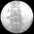
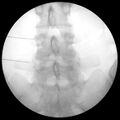 MBB Left L3 and L4.jpg 1,162 × 1,162; 139 KB
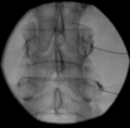
 MBB Left L3 and L4 AP contrast.jpg 1,162 × 1,162; 115 KB
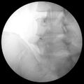
 MBB Left L3 and L4 Decline.jpg 1,162 × 1,162; 144 KB
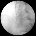
 MBB Left L3 and L4 Oblique.jpg 1,162 × 1,162; 141 KB
 MBB facet joint.png 403 × 397; 31 KB
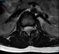 MRI T2 Lumbar Spine L1-L2 Transarticular Axial.jpg 990 × 900; 134 KB
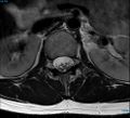 MRI T2 Lumbar Spine L1 Subpedicular Axial.jpg 990 × 900; 133 KB
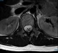 MRI T2 Lumbar Spine L1 Transpedicular Axial.jpg 990 × 900; 131 KB


