File:Cor T2 MRI tropical pyomyositis.jpg: Difference between revisions
From WikiMSK
(Coronal T2 weighted fat suppressed image showing a multiloculated fluid collection in the left gluteal musculature due to tropical pyomositis in a 12 year old boy. From https://commons.wikimedia.org/wiki/File:Cor_T2_MRI_tropical_pyomyositis.JPG) |
(No difference)
|
Latest revision as of 22:04, 9 April 2022
Summary
Coronal T2 weighted fat suppressed image showing a multiloculated fluid collection in the left gluteal musculature due to tropical pyomositis in a 12 year old boy. From https://commons.wikimedia.org/wiki/File:Cor_T2_MRI_tropical_pyomyositis.JPG
Licencing
![]()
This work is licensed under the Creative Commons Attribution-ShareAlike 4.0 International License.
File history
Click on a date/time to view the file as it appeared at that time.
| Date/Time | Thumbnail | Dimensions | User | Comment | |
|---|---|---|---|---|---|
| current | 22:04, 9 April 2022 | 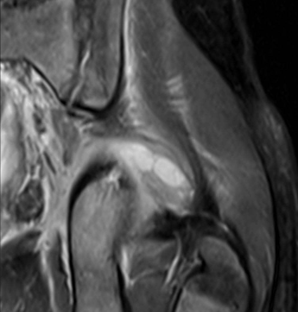 | 1,004 × 1,050 (60 KB) | Jeremy (talk | contribs) | Coronal T2 weighted fat suppressed image showing a multiloculated fluid collection in the left gluteal musculature due to tropical pyomositis in a 12 year old boy. From https://commons.wikimedia.org/wiki/File:Cor_T2_MRI_tropical_pyomyositis.JPG |
You cannot overwrite this file.
File usage
The following file is a duplicate of this file (more details):
- File:Cor T2 MRI tropical pyomyositis.JPG from Wikimedia Commons
The following page uses this file:

